. 2012 Apr 10;14(2):R59.
doi: 10.1186/bcr3163.

Characterizing mammographic images by using generic texture features

Affiliations

Characterizing mammographic images by using generic texture features

Lothar Häberle et al. Breast Cancer Res. .

Abstract

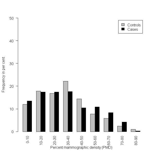
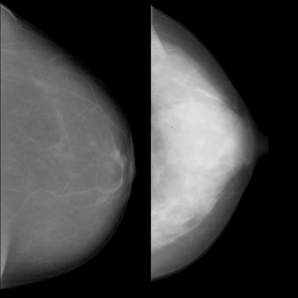
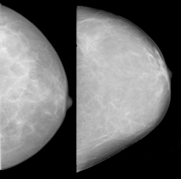
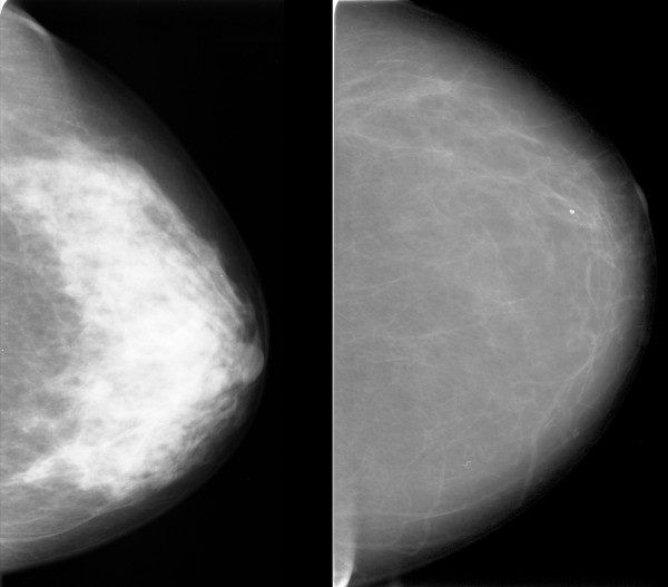
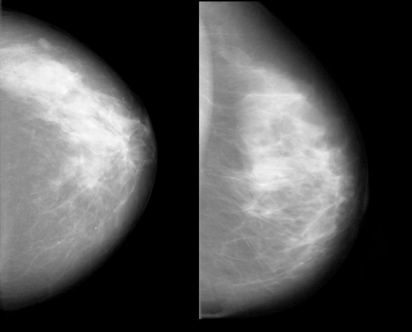
Introduction: Although mammographic density is an established risk factor for breast cancer, its use is limited in clinical practice because of a lack of automated and standardized measurement methods. The aims of this study were to evaluate a variety of automated texture features in mammograms as risk factors for breast cancer and to compare them with the percentage mammographic density (PMD) by using a case-control study design.

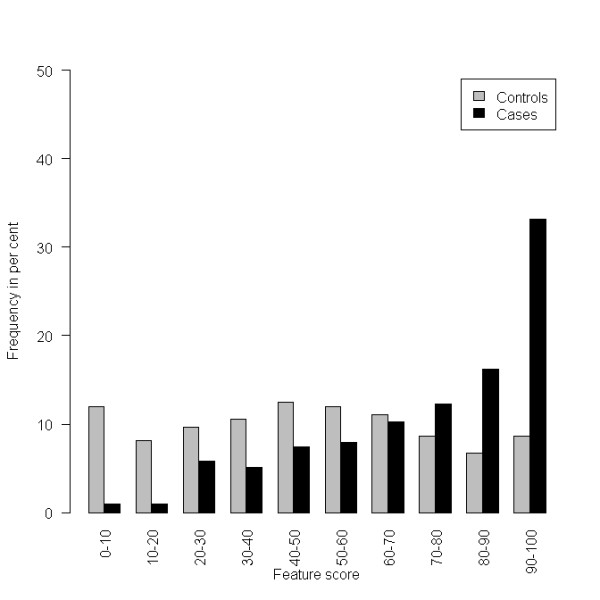
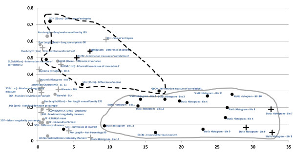
Methods: A case-control study including 864 cases and 418 controls was analyzed automatically. Four hundred seventy features were explored as possible risk factors for breast cancer. These included statistical features, moment-based features, spectral-energy features, and form-based features. An elaborate variable selection process using logistic regression analyses was performed to identify those features that were associated with case-control status. In addition, PMD was assessed and included in the regression model.

Results: Of the 470 image-analysis features explored, 46 remained in the final logistic regression model. An area under the curve of 0.79, with an odds ratio per standard deviation change of 2.88 (95% CI, 2.28 to 3.65), was obtained with validation data. Adding the PMD did not improve the final model.

Conclusions: Using texture features to predict the risk of breast cancer appears feasible. PMD did not show any additional value in this study. With regard to the features assessed, most of the analysis tools appeared to reflect mammographic density, although some features did not correlate with PMD. It remains to be investigated in larger case-control studies whether these features can contribute to increased prediction accuracy.


Figures

Figure 1
Figure 1
Histogram of the final feature score, based on the 46 finally selected features applied on the validation data set.
Figure 2
Figure 2
Histogram of the percentage mammographic density (PMD) on the validation data set.
Figure 3
Figure 3
Finally selected features (n = 46). Strength of risk prediction within the final logistic regression model on x-axis (absolute value of log odds ratio per standard deviation) and the feature's Spearman correlation with percentage mammographic density (PMD) on the y-axis. +The texture feature and PMD have the same direction with regard to their association with risk (that is, either positive log OR and positive correlation with PMD or negative log OR and negative correlation with PMD). The texture feature and PMD have the opposite direction with regard to their association with risk (that is, either positive log OR and negative correlation or negative log OR and positive correlation with PMD). Gray symbols, Feature is selected in fewer than 90% of the bootstrap samples. Black symbols, It is selected in more than 90% of the bootstrap samples. The dashed line circumscribes a cluster of second-order statistical features, and the continuous gray line circumscribes a cluster of first-order statistical features. "Static histogram" refers to features describing the relative frequency of gray-level values according to a given interval (bin). These features are thus first-order statistics describing the gray-level distribution. SDH refers to features calculated from sum and difference histograms, and GLCM refers to features calculated from a gray level co-occurrence matrix. Both of these are second-order statistics, describing the gray-level distribution relative to spatial relations between adjacent pixels. SGF refers to the statistical geometric features, describing the structure of the microtexture. A more-detailed description of all of the features is given in the Methods section.
Figure 4
Figure 4
Example of a feature with the same direction for the correlation of the feature with breast cancer risk and percentage mammographic density (PMD). Patients with mammograms like that on the left had low values for the feature "SDH (0.5 cm) difference of contrast" and had a low predicted risk of breast cancer. Patients with mammograms like that on the right had high feature values, a high risk of breast cancer, and a high mammographic density. The Spearman correlation with PMD for this feature was +0.54.
Figure 5
Figure 5
Example of a feature with no correlation with percentage mammographic density (PMD). Patients with mammograms like that on the left had low values for the feature "GLCM inverse difference moment" and had a low predicted risk of breast cancer. Patients with mammograms like that on the right had high feature values and a high risk of breast cancer. The Spearman correlation with PMD for this feature was -0.05.
Figure 6
Figure 6
Example of a feature with different directions for the correlation with breast cancer risk and PMD. Patients with mammograms like that on the left had low values for the feature "SDH (0.5 cm) difference of entropies" and had a low predicted risk of breast cancer and a high mammographic density. Patients with mammograms like that on the right had high feature values, a high risk of breast cancer, and a low mammographic density. The Spearman correlation with PMD for this feature was -0.72.
Figure 7
Figure 7
Examples of images with low score values calculated with the final prediction model and a low risk of breast cancer (left), and images with high score values and a high risk of breast cancer (right). Spearman's rho for the correlation between the final score and percentage mammographic density (PMD) was 0.02.

References

    1. Boyd NF, Guo H, Martin LJ, Sun L, Stone J, Fishell E, Jong RA, Hislop G, Chiarelli A, Minkin S, Yaffe MJ. Mammographic density and the risk and detection of breast cancer. N Engl J Med. 2007;356:227–236. doi: 10.1056/NEJMoa062790. - DOI - PubMed
    1. McCormack VA, dos Santos Silva I. Breast density and parenchymal patterns as markers of breast cancer risk: a meta-analysis. Cancer Epidemiol Biomarkers Prev. 2006;15:1159–1169. doi: 10.1158/1055-9965.EPI-06-0034. - DOI - PubMed
    1. Fasching PA, Ekici AB, Adamietz BR, Wachter DL, Hein A, Bayer CM, Häberle L, Loehberg CR, Jud SM, Heusinger K, Rubner M, Rauh C, Bani MR, Lux MP, Schulz-Wendtland R, Hartmann A, Beckmann MW. Breast cancer risk: genes, environment and clinics. Geburtsh Frauenheilk. 2011;71:1056–1066. - PMC - PubMed
    1. Wolfe JN. Breast patterns as an index of risk for developing breast cancer. AJR Am J Roentgenol. 1976;126:1130–1137. - PubMed
    1. Wolfe JN, Saftlas AF, Salane M. Mammographic parenchymal patterns and quantitative evaluation of mammographic densities: a case-control study. AJR Am J Roentgenol. 1987;148:1087–1092. - PubMed

Publication types