

Toward routine use of 3D histopathology as a research tool
- PMID: 22490922
- PMCID: PMC3538002
- DOI: 10.1016/j.ajpath.2012.01.033
Toward routine use of 3D histopathology as a research tool
Erratum in
- Am J Pathol. 2012 Jul;181(1):374
Abstract
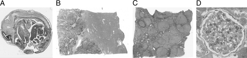
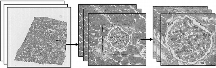
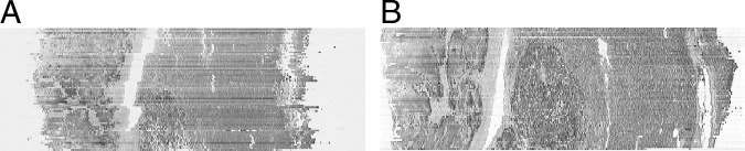
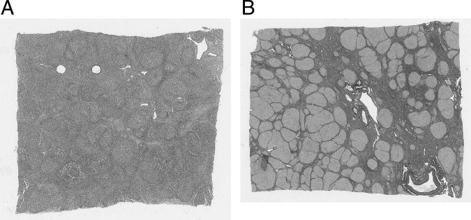
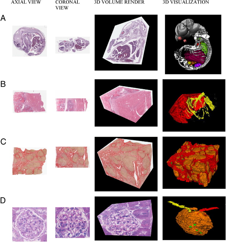
Three-dimensional (3D) reconstruction and examination of tissue at microscopic resolution have significant potential to enhance the study of both normal and disease processes, particularly those involving structural changes or those in which the spatial relationship of disease features is important. Although other methods exist for studying tissue in 3D, using conventional histopathological features has significant advantages because it allows for conventional histopathological staining and interpretation techniques. Until now, its use has not been routine in research because of the technical difficulty in constructing 3D tissue models. We describe a novel system for 3D histological reconstruction, integrating whole-slide imaging (virtual slides), image serving, registration, and visualization into one user-friendly package. It produces high-resolution 3D reconstructions with minimal user interaction and can be used in a histopathological laboratory without input from computing specialists. It uses a novel method for slice-to-slice image registration using automatic registration algorithms custom designed for both virtual slides and histopathological images. This system has been applied to >300 separate 3D volumes from eight different tissue types, using a total of 5500 virtual slides comprising 1.45 TB of primary image data. Qualitative and quantitative metrics for the accuracy of 3D reconstruction are provided, with measured registration accuracy approaching 120 μm for a 1-cm piece of tissue. Both 3D tissue volumes and generated 3D models are presented for four demonstrator cases.
Copyright © 2012 American Society for Investigative Pathology. Published by Elsevier Inc. All rights reserved.
Figures
References
-
- Kaufman M.H., Brune R.M., Baldock R.A., Bard J.B.L., Davidson D. Computer-aided 3D reconstruction of serially sectioned mouse embryos: its use in integrating anatomical organization. Int J Dev Biol. 1997;41:223–233. - PubMed
-
- Wang X., Lindsay S., Baldock R. From spatial-data to 3D models of the developing human brain. Methods. 2010;50:96–104. - PubMed
-
- Quintana L., Sharpe J. Optical projection tomography of vertebrate embryo development. Cold Spring Harb Protoc. 2011;2011:586–594. - PubMed
-
- Prager R.W., Ijaz U.Z., Gee A.H., Treece G.M. Three-dimensional ultrasound imaging. Proc Inst Mech Eng H. 2010;224:193–223. - PubMed
Publication types
MeSH terms
Grants and funding
LinkOut - more resources
Full Text Sources
Other Literature Sources
