. 2012 May 15;188(10):4876-84.
doi: 10.4049/jimmunol.1200389. Epub 2012 Apr 9.

PD-1 protects against inflammation and myocyte damage in T cell-mediated myocarditis

Affiliations

PD-1 protects against inflammation and myocyte damage in T cell-mediated myocarditis

Margarite L Tarrio et al. J Immunol. .

Abstract

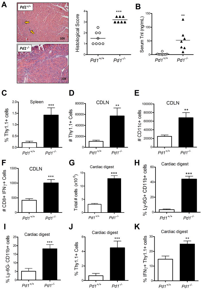
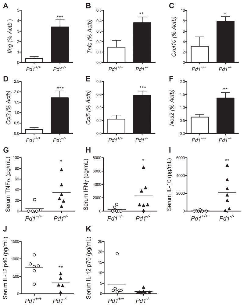
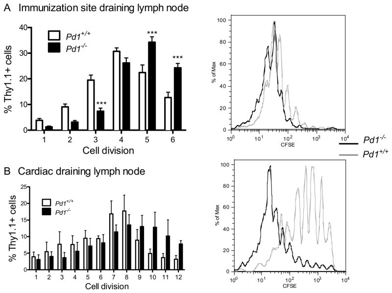
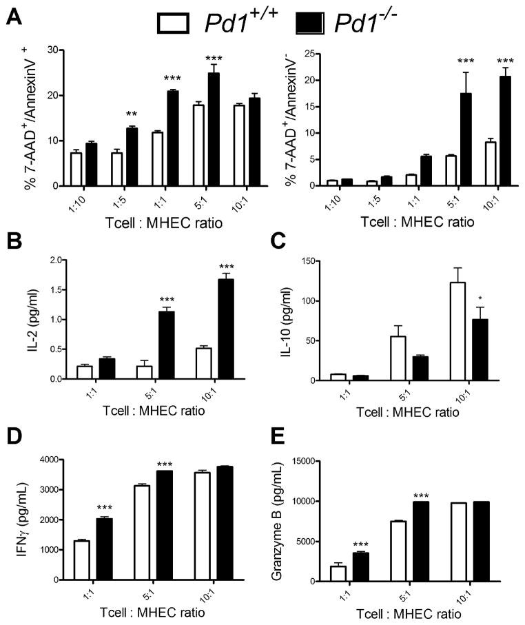
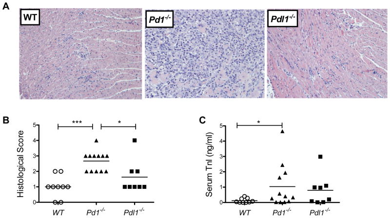
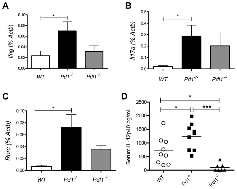
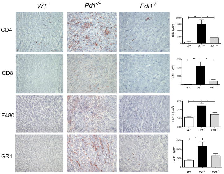
PD-1, a member of the CD28 family of immune regulatory molecules, is expressed on activated T cells, interacts with its ligands, PD-L1/B7-H1 and PD-L2/B7-DC, on other cells, and delivers inhibitory signals to the T cell. We studied the role of this pathway in modulating autoreactive T cell responses in two models of myocarditis. In a CD8(+) T cell-mediated adoptive transfer model, we found that compared with Pd1(+/+) CD8(+) T cells, Pd1(-/-) CD8(+) T cells cause enhanced disease, with increased inflammatory infiltrate, particularly rich in neutrophils. Additionally, we show enhanced proliferation in vivo and enhanced cytotoxic activity of PD-1-deficient T lymphocytes against myocardial endothelial cells in vitro. In experimental autoimmune myocarditis, a disease model dependent on CD4(+) T cells, we show that mice lacking PD-1 develop enhanced disease compared with wild-type mice. PD-1-deficient mice displayed increased inflammation, enhanced serum markers of myocardial damage, and an increased infiltration of inflammatory cells, including CD8(+) T cells. Together, these studies show that PD-1 plays an important role in limiting T cell responses in the heart.


Figures

Figure 1
Figure 1. PD-1 controls pathologic CD8+ T cell responses in the heart
Naive WT or PD-1 deficient CD8+ OT-1+ T cells were adoptively transferred into cMy-mOVA mice, immunized 24 hours later with CFA/Ovalbumin, and sacrificed 7 days later. Whole heart H&E sections were prepared and scored for myocarditis (A), and circulating troponin was measured in the serum (B). Flow cytometric analysis was performed for identification of transferred cells in the spleen (C), and cardiac draining lymph node (D), as well as for identification of dendritic cells (E), and IFNγ-producing cells (F), in draining lymph node. Enzymatic digestion of whole hearts was performed and total cell number (G), infiltrating neutrophils (H), infiltrating monocytes (I), infiltrating transferred OT-1 cells (J), and IFNγ-producing transferred cells (K), were enumerated by flow cytometry. In A and B data are pooled form three experiments, each one with its own Pd1+/+ and Pd1−/− OT-1 T cell preparations. Each symbol represent the mean of replicate measurements of individual mice, and the horizontal bars are the S.E.M. of all the mice. Data in C-K represent mean ± S.E.M, N=3 mice, of samples from one of the three experiments,. * P<0.05, ** P<0.01 *** P< 0.001.
Figure 2
Figure 2. PD-1 regulates pathogenic gene expression in the heart, and influences circulating pathogenic cytokines
RNA was purified from heart tissue of the mice described in Figure 1 and used for qRT-PCR analysis of Ifng, Tnfa, Cxcl10, Ccl3, Ccl5, and Nos2 (A–F). Serum samples from the time of sacrifice were analyzed using multiplex luminex-bead based cytokine assays for TNFα, IFNγ, IL-10, IL-12p40, and IL-12p70 (G–K). Data are the mean ± S.E.M. N=3 mice. * P<0.05, ** P<0.01 *** P< 0.001
Figure 3
Figure 3. PD-1 controls in vivo proliferation of transferred T cells
Naïve WT or PD-1 deficient OT-1+ CD8+ T cells were labeled with CFSE, and then transferred into cMy-mOVA recipients. The recipient mice immunized s.c. 24 hours later with CFA/Ovalbumin, and sacrificed 72 hours later. Immunization site draining lymph nodes (A), and cardiac draining lymph nodes (B), were analyzed for CFSE dilution of Thy1.1+ transferred cells. Data presented represent the mean ± S.E.M. N=3 mice, from one of 3 experiments with similar results. * P<0.05, ** P<0.01 *** P< 0.001
Figure 4
Figure 4. PD-1 deficient CTLs are more effective killers of cardiac-derived target cells, in vitro
Naïve OT-1+ CD8+ T cells were isolated from Pd1−/− or WT mice, and cultured for 5 days in the presence of IL-2 and IL-12 to generate effector cells, and then rested overnight. Confluent mouse heart endothelial cells (MHEC) were pre-treated with IFNγ for 2 h and pulsed with the ovalbumin peptide SIINFKL. The MHEC were then co-cultured with OT-1 effectors at the indicated ratios of effector to target cell ratios for 1 h, and were then analyzed by FACS for Annexin V and 7-AAD staining (A). Supernatants from these assays were analyzed for the presence of IL-2 (B) and IL-10 (C) using multiplex luminex-bead based cytokine assays, and IFNγ (D) and granzyme B (E) by ELISA. Data presented represent the mean ± S.E.M of 3 experiments. * P<0.05, ** P<0.01 *** P< 0.001
Figure 5
Figure 5. PD-1 deficiency exacerbates experimental autoimmune myocarditis
WT, Pd1−/− or Pdl1−/− BALB/c mice were immunized at day 0 and day 7 with CFA/γ-myosin heavy chain peptide, and sacrificed at day 21. Representative sections of H&E stained sections of heart tissue are shown (A). Sections were scored for histopathology (B), and troponin I levels were measured in serum collected at sacrifice (C). Data are mean ± S.E.M. of 7–10 mice per group. * P<0.05, ** P<0.01 *** P< 0.001
Figure 6
Figure 6. PD-1 regulates cardiac inflammatory gene expression in EAM and the release of circulating cytokines
RNA was isolated from heart tissue of the mice described in Figure 5, sampled at the time of sacrifice, and analyzed by qRT-PCR for expression of Ifng(A), Il17α (B), and Rorc (B). Serum samples from day 14 and day 21 were analyzed using multiplex luminex-bead based cytokine assays for the presence of IL-12p40 (D). Data are the mean ± S.E.M. * P<0.05, ** P<0.01 *** P< 0.001
Figure 7
Figure 7. PD-1 deficiency results in increased inflammatory infiltrates EAM
Immunohistochemical analysis was performed in heart sections of the mice described in Figures 5 and 6 for CD4, CD8, F480 (macrophages), and Ly6G (neutrophils). Total positive staining area was quantified using ImagePro softwareJ. Error bar represent the mean ± S.E.M. * P<0.05, ** P<0.01 *** P< 0.001

References

    1. Rose NR, Beisel KW, Herskowitz A, Neu N, Wolfgram LJ, Alvarez FL, Traystman MD, Craig SW. Cardiac myosin and autoimmune myocarditis. Ciba Found Symp. 1987;129:3–24. - PubMed
    1. Cihakova D, Rose NR. Pathogenesis of myocarditis and dilated cardiomyopathy. Adv Immunol. 2008;99:95–114. - PubMed
    1. Baldeviano GC, Barin JG, Talor MV, Srinivasan S, Bedja D, Zheng D, Gabrielson K, Iwakura Y, Rose NR, Cihakova D. Interleukin-17A is dispensable for myocarditis but essential for the progression to dilated cardiomyopathy. Circ Res. 2010;106:1646–1655. - PubMed
    1. Sonderegger I, Rohn TA, Kurrer MO, Iezzi G, Zou Y, Kastelein RA, Bachmann MF, Kopf M. Neutralization of IL-17 by active vaccination inhibits IL-23-dependent autoimmune myocarditis. Eur J Immunol. 2006;36:2849–2856. - PubMed
    1. Afanasyeva M, Georgakopoulos D, Rose NR. Autoimmune myocarditis: cellular mediators of cardiac dysfunction. Autoimmun Rev. 2004;3:476–486. - PubMed

Publication types

MeSH terms

Substances