

Culturing and applications of rotating wall vessel bioreactor derived 3D epithelial cell models
- PMID: 22491366
- PMCID: PMC3567125
- DOI: 10.3791/3868
Culturing and applications of rotating wall vessel bioreactor derived 3D epithelial cell models
Abstract
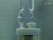
Cells and tissues in the body experience environmental conditions that influence their architecture, intercellular communications, and overall functions. For in vitro cell culture models to accurately mimic the tissue of interest, the growth environment of the culture is a critical aspect to consider. Commonly used conventional cell culture systems propagate epithelial cells on flat two-dimensional (2-D) impermeable surfaces. Although much has been learned from conventional cell culture systems, many findings are not reproducible in human clinical trials or tissue explants, potentially as a result of the lack of a physiologically relevant microenvironment. Here, we describe a culture system that overcomes many of the culture condition boundaries of 2-D cell cultures, by using the innovative rotating wall vessel (RWV) bioreactor technology. We and others have shown that organotypic RWV-derived models can recapitulate structure, function, and authentic human responses to external stimuli similarly to human explant tissues (1-6). The RWV bioreactor is a suspension culture system that allows for the growth of epithelial cells under low physiological fluid shear conditions. The bioreactors come in two different formats, a high-aspect rotating vessel (HARV) or a slow-turning lateral vessel (STLV), in which they differ by their aeration source. Epithelial cells are added to the bioreactor of choice in combination with porous, collagen-coated microcarrier beads (Figure 1A). The cells utilize the beads as a growth scaffold during the constant free fall in the bioreactor (Figure 1B). The microenvironment provided by the bioreactor allows the cells to form three-dimensional (3-D) aggregates displaying in vivo-like characteristics often not observed under standard 2-D culture conditions (Figure 1D). These characteristics include tight junctions, mucus production, apical/basal orientation, in vivo protein localization, and additional epithelial cell-type specific properties. The progression from a monolayer of epithelial cells to a fully differentiated 3-D aggregate varies based on cell type(1, 7-13). Periodic sampling from the bioreactor allows for monitoring of epithelial aggregate formation, cellular differentiation markers and viability (Figure 1D). Once cellular differentiation and aggregate formation is established, the cells are harvested from the bioreactor, and similar assays performed on 2-D cells can be applied to the 3-D aggregates with a few considerations (Figure 1E-G). In this work, we describe detailed steps of how to culture 3-D epithelial cell aggregates in the RWV bioreactor system and a variety of potential assays and analyses that can be executed with the 3-D aggregates. These analyses include, but are not limited to, structural/morphological analysis (confocal, scanning and transmission electron microscopy), cytokine/chemokine secretion and cell signaling (cytometric bead array and Western blot analysis), gene expression analysis (real-time PCR), toxicological/drug analysis and host-pathogen interactions. The utilization of these assays set the foundation for more in-depth and expansive studies such as metabolomics, transcriptomics, proteomics and other array-based applications. Our goal is to present a non-conventional means of culturing human epithelial cells to produce organotypic 3-D models that recapitulate the human in vivo tissue, in a facile and robust system to be used by researchers with diverse scientific interests.
References
-
- Herbst-Kralovetz MM, et al. Quantification and comparison of toll-like receptor expression and responsiveness in primary and immortalized human female lower genital tract epithelia. Am. J. Reprod. Immunol. 2008;59(3):212–224. - PubMed
-
- Bibliography of Research Publications [Internet] Houston, Texas: Synthecon; 2012. [cited 2001 Mar 8]. Available from: http://www.synthecon.com/bibliography.html.
-
- Khaoustov VI, et al. Induction of three-dimensional assembly of human liver cells by simulated microgravity. In Vitro Cell Dev. Biol. Anim. 1999;35:501–509. - PubMed
-
- Papadaki M, et al. Tissue engineering of functional cardiac muscle: molecular, structural, and electrophysiological studies. Am. J. Physiol. Heart Circ. Physiol. 2001;280:168–178. - PubMed
Publication types
MeSH terms
Grants and funding
LinkOut - more resources
Full Text Sources
Other Literature Sources
