. 2012 Jun;86(12):6434-43.
doi: 10.1128/JVI.00433-12. Epub 2012 Apr 4.

A 3-O-sulfated heparan sulfate binding peptide preferentially targets herpes simplex virus 2-infected cells

Affiliations

A 3-O-sulfated heparan sulfate binding peptide preferentially targets herpes simplex virus 2-infected cells

Mohamed M Ali et al. J Virol. 2012 Jun.

Abstract

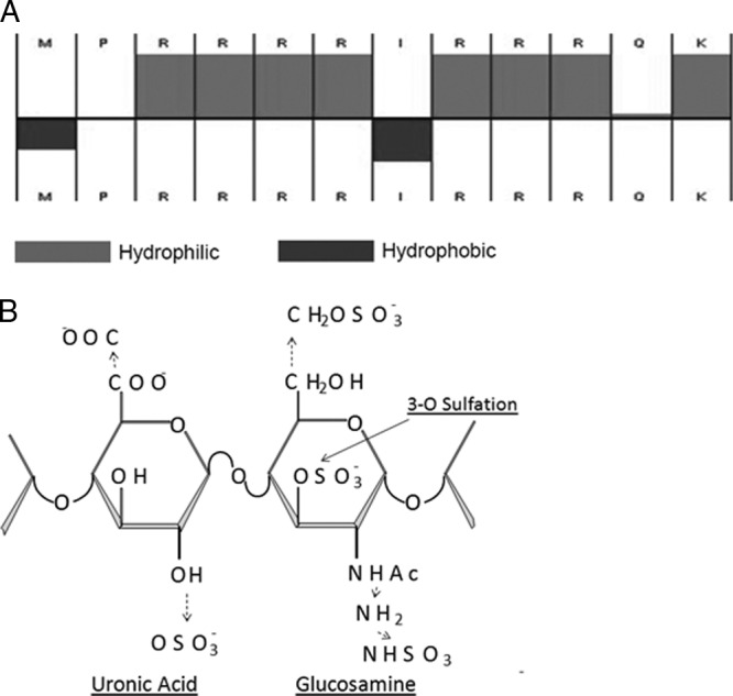
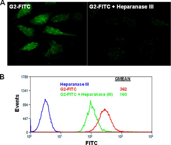
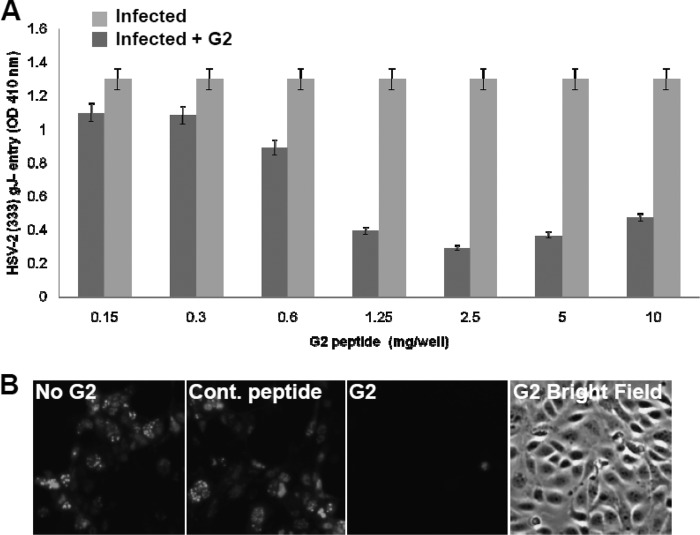
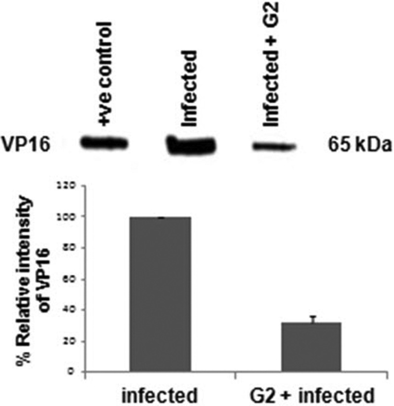
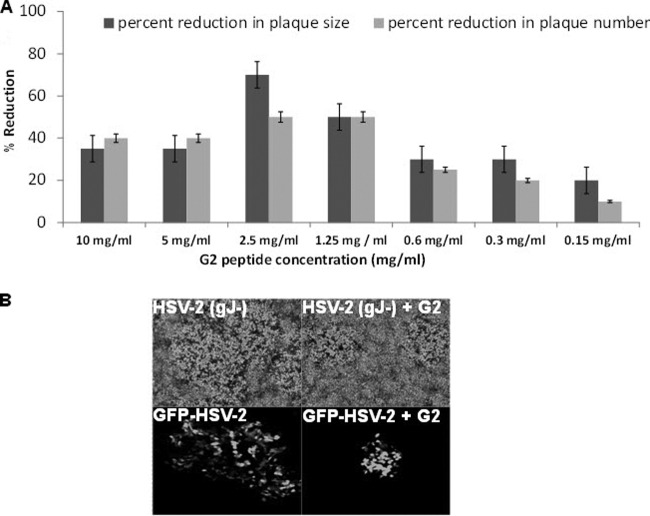
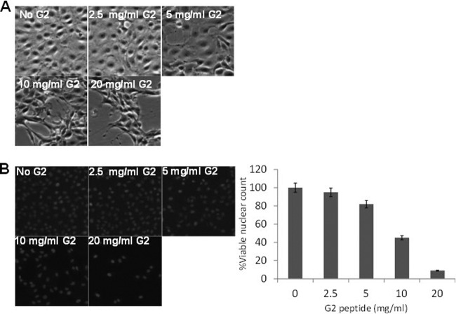
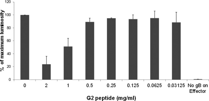
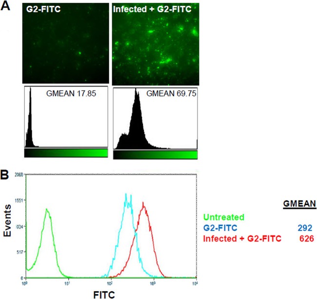
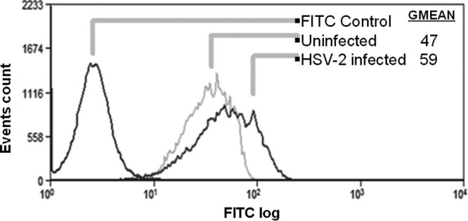
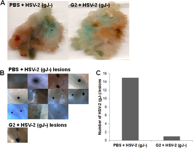
Herpes simplex virus 2 (HSV-2) is the primary cause of genital herpes, which is one of the most common sexually transmitted viral infections worldwide and a major cofactor for human immunodeficiency virus infection. The lack of an effective vaccine or treatment and the emergence of drug-resistant strains highlight the need for developing new antivirals for HSV-2. Here, we demonstrate that a low-molecular-weight peptide isolated against 3-O-sulfated heparan sulfate (3-OS HS) can efficiently block HSV-2 infection. Treatment with the peptide inhibited viral entry and cell-to-cell spread both in vitro and in vivo using a mouse model of genital HSV-2 infection. Quite interestingly, the peptide showed a preferential binding to HSV-2-infected cells, with more than 200% increased binding compared to uninfected cells. Our additional results show that heparan sulfate expression is upregulated by 25% upon HSV-2 infection, which is a significant new finding that could be exploited for designing new diagnostic tests and treatment strategies against HSV-2-infected cells. In addition, our results also raise the possibility that 3-OS HS modifications within HS may be upregulated even more to accommodate for a significantly higher increase in the peptide binding to the infected cells.


Figures

Fig 1
Fig 1
G2 peptide structure. (A) Arrangement of the hydrophilic (gray) and hydrophobic (black) amino acid residues of the G2 peptide as calculated by Innovagen peptide property calculator (Hopp & Woods), accessible from http://www.innovagen.se/custom-peptide-synthesis/peptide-property-calculator/peptide-property-calculator.asp (as of August 2011). (B) 3-O-Sulfated HS. The disaccharide structure representative of 3-O-sulfated HS is shown. The solid arrow shows the 3-O position of the glucosamine residue where sulfation is essential for HSV-1 gD binding. The broken arrows show the additional modifications, one or more of which may also be present as part of the gD-binding 3-OS HS.
Fig 2
Fig 2
G2 peptide binds specifically to HS. (A) FITC-conjugated G2 peptide (FITC-G2) binding to Vero cells with or without heparanase III treatment imaged by fluorescence microscopy. Vero cells were grown in chamber slides and either treated or not treated with heparanase III (10 mIU) for 2 h at 37°C. This was followed by incubation at 37°C for 30 min with FITC-G2 peptide. (B) FITC-G2 peptide binding to Vero cells with or without heparanase III treatment quantified by flow cytometry. Vero cells were treated as described for panel A and processed for flow cytometry. Vero cells treated with heparanase III only were used as a background control. Results are representative of three independent experiments.
Fig 3
Fig 3
G2 peptide inhibits HSV-2 entry into HeLa cells. (A) Confluent cultures of HeLa cells in a 96-well plate were either left untreated or treated with serial dilutions of G2 peptide for 1 h at 37°C. Cells were then infected with the recombinant β-galactosidase-expressing HSV-2 strain 333 (gJ) at an MOI of 20. After 6 h, the soluble substrate ONPG was added and enzymatic activity was measured. Results are representative of three independent experiments. (B) The effect of entry-blocking activity of G2 peptide was examined by fluorescence microscopy. Vero cells were not treated (No G2), treated with control nonspecific peptide (Cont. peptide), or treated with G2 peptide (G2) at 1 mg/ml for 1 h. Cells were then infected with GFP-expressing HSV-2 (GFP–HSV-2) at an MOI of 10 for 2 h, washed, and incubated for another 12 h. Imaging was performed using a ×10 objective on a Zeiss Axiovert 200 fluorescence microscope. Representative images from one experiment performed in triplicate are shown. Images of G2 peptide-treated cells under both the fluorescent light and the bright field are shown.
Fig 4
Fig 4
G2 peptide pretreatment reduces VP16 protein translocation into cells. As a marker for HSV-2 entry, the VP16 level was measured by Western blotting using lysates from HeLa cells untreated or treated with G2 peptide (2 mg/ml) for 30 min, followed by HSV-2 infection for 4 h. A representative Western blot is shown. The positive control represents lysates from cells transiently expressing VP16. The results are expressed as mean ± 1 SD values from two independent experiments.
Fig 5
Fig 5
The IC50 of G2 peptide is approximately 1 mg/ml by plaque reduction assay. Vero cells were either untreated or treated with serial dilutions of G2 peptide for 1 h at 37°C. Cells were then infected at 20 to 30 PFU/well with either HSV-2(333) gJ or GFP-tagged HSV-2 (GFP–HSV-2) for 2 h at 37°C. After 2 h, cells were washed and overlaid with 1% methylcellulose in DMEM supplemented with 0.05% human pooled IgG for 48 to 72 h. (A) HSV-2 plaque number and size. Plaque number was counted under the microscope. Plaque size was measured using the Axiovision software, version 4, program. Results are representative of three independent experiments. (B) Representative images of HSV-2(333) gJ plaques after crystal violet staining (top) and GFP–HSV-2 plaques under fluorescent excitation light (bottom). Imaging was performed using a Zeiss Axioscope microscope equipped with a digital low-light CCD camera under the control of the imaging software Axiovision at a ×100 magnification.
Fig 6
Fig 6
G2 peptide affects cell viability at concentrations above 2.5 mg/ml in a dose-dependent manner. (A) HCE cells were either untreated or treated with serial dilutions of G2 peptide in MEM complete medium for 24 h at 37°C in 5% CO2. The morphological appearance of treated HCE cells was observed and compared to that of untreated cells at ×100 magnification. (B) Viability of Vero cells that were either untreated or treated with serial dilutions of G2 peptide in DMEM complete medium for 24 h at 37°C in 5% CO2 was examined by counting fluorescent nuclei of viable cells after staining the cells with Hoechst 33342 live-cell nuclear stain. Representative images of each condition are shown, as is the relative number of viable cells. Results are representative of three independent experiments.
Fig 7
Fig 7
Pretreatment of target cells with G2 peptide affects cell-to-cell fusion in a dose-dependent manner. Target CHO-K1 cells expressing HSV-2 gD receptor nectin-1 and the luciferase gene under the control of the T7 promoter were preincubated with a serial dilution of G2 peptide or left untreated for 1 h at 37°C. Target cells were then mixed with an effector cell population that expresses HSV-1 fusion glycoproteins plus T7 polymerase. As a negative control for the cell fusion assay, untreated target cells were mixed with effector cells that lack HSV-2 gB. Luciferase reporter gene activity was determined to quantify cell-to-cell fusion. Results are presented as mean ± 1 SD of 3 independent experiments.
Fig 8
Fig 8
G2 peptide is preferentially bound by an infected cell population. Vero cells were either uninfected or infected with HSV-2 at an MOI of 10 for 2 h at 37°C. Cells were then treated with 1 mg/ml FITC-conjugated G2 peptide (G2-FITC) for 30 min at 37°C. (A) G2-FITC binding was examined by fluorescence microscopy at ×100 magnification. Images were analyzed using the imageJ histogram analysis function. (B) G2-FITC binding by uninfected or HSV-2-infected cells was further quantified using flow cytometry analysis. Results are representative of six independent experiments.
Fig 9
Fig 9
HSV-2 infection results in an increase in HS expression on the surface of infected Vero cells. The effect of HSV-2 infection on HS expression was analyzed using flow cytometric analysis of HS expression probed by FITC-conjugated anti-HS antibody specific to epitope 10E4. Vero cells were infected with HSV-2 strain 333 at an MOI of 10 for 2 h. Vero cells incubated with a FITC-conjugated secondary antibody (isotypic control) were used as a background control. Results are representative of three independent experiments.
Fig 10
Fig 10
Intravaginal G2-peptide pretreatment reduces the number of HSV-2-infected cell colonies. BALB/c female mice were pretreated for 1 h with either PBS or 25 μl G2 peptide (2 mg/ml). Mice were then infected with 1 × 10³ PFU β-galactosidase-expressing HSV-2 (gJ). At 24 h after infection, mice were sacrificed and the vaginal tissue was harvested, fixed, and incubated in X-Gal in ferricyanide buffer to stain infected cell colonies. Tissue samples were compressed onto a glass slide and imaged using a Zeiss Axioscope microscope equipped with a digital camera at ×100 magnification. (A) Vaginal tissue from mice of the indicated treatments; (B) HSV-2 (gJ) vaginal lesions found in the vaginas from mice for the treatments noted; (C) quantitation of the vaginal lesions.

References

    1. Akhtar J, et al. 2008. HVEM and nectin-1 are the major mediators of herpes simplex virus 1 (HSV-1) entry into human conjunctival epithelium. Invest. Ophthalmol. Vis. Sci. 49:4026–4035 - PMC - PubMed
    1. Albiol Matanic VC, Castilla V. 2004. Antiviral activity of antimicrobial cationic peptides against Junin virus and herpes simplex virus. Int. J. Antimicrob. Agents 23:382–389 - PubMed
    1. Andersen JH, Jenssen H, Gutteberg TJ. 2003. Lactoferrin and lactoferricin inhibit herpes simplex 1 and 2 infection and exhibit synergy when combined with acyclovir. Antiviral Res. 58:209–215 - PubMed
    1. Andersen JH, Jenssen H, Sandvik K, Gutteberg TJ. 2004. Anti-HSV activity of lactoferrin and lactoferricin is dependent on the presence of heparan sulphate at the cell surface. J. Med. Virol. 74:262–271 - PubMed
    1. Araki-Sasaki K, et al. 1995. An SV40-immortalized human corneal epithelial cell line and its characterization. Invest. Ophthalmol. Vis. Sci. 36:614–621 - PubMed

Publication types