. 2012 Jun;86(12):6792-803.
doi: 10.1128/JVI.07172-11. Epub 2012 Apr 4.

Multifunctional CD4 cells expressing gamma interferon and perforin mediate protection against lethal influenza virus infection

Affiliations

Multifunctional CD4 cells expressing gamma interferon and perforin mediate protection against lethal influenza virus infection

Deborah M Brown et al. J Virol. 2012 Jun.

Abstract

CD4 effectors generated in vitro can promote survival against a highly pathogenic influenza virus via an antibody-independent mechanism involving class II-restricted, perforin-mediated cytotoxicity. However, it is not known whether CD4 cells activated during influenza virus infection can acquire cytolytic activity that contributes to protection against lethal challenge. CD4 cells isolated from the lungs of infected mice were able to confer protection against a lethal dose of H1N1 influenza virus A/Puerto Rico 8/34 (PR8). Infection of BALB/c mice with PR8 induced a multifunctional CD4 population with proliferative capacity and ability to secrete interleukin-2 (IL-2) and tumor necrosis factor alpha (TNF-α) in the draining lymph node (DLN) and gamma interferon (IFN-γ) and IL-10 in the lung. IFN-γ-deficient CD4 cells produced larger amounts of IL-17 and similar levels of TNF-α, IL-10, and IL-2 compared to wild-type (WT) CD4 cells. Both WT and IFN-γ(-/-) CD4 cells exhibit influenza virus-specific cytotoxicity; however, IFN-γ-deficient CD4 cells did not promote recovery after lethal infection as effectively as WT CD4 cells. PR8 infection induced a population of cytolytic CD4 effectors that resided in the lung but not the DLN. These cells expressed granzyme B (GrB) and required perforin to lyse peptide-pulsed targets. Lethally infected mice given influenza virus-specific CD4 cells deficient in perforin showed greater weight loss and a slower time to recovery than mice given WT influenza virus-specific CD4 cells. Taken together, these data strengthen the concept that CD4 T cell effectors are broadly multifunctional with direct roles in promoting protection against lethal influenza virus infection.


Figures

Fig 1
Fig 1
CD4 effectors from the DLN and lung provide protection from lethal-dose influenza virus infection. Healthy BALB/c mice were infected with 500 EIU PR8 i.n., and 7 to 10 days later, CD4 cells were isolated from the DLN or lung using anti-CD4-conjugated magnetic beads. CD4 cells from the DLN (A), lung (B), or DLN and lung combined (C) were adoptively transferred intravenously to naïve BALB/c mice that were subsequently infected with 5,000 EIU PR8, and survival was monitored over time. Kaplan-Meier curves were generated by Prism software. In all graphs, survival of mice given CD4 cells is significantly higher than that in mice left untreated (none) by the log-rank test (P ≤ 0.01).
Fig 2
Fig 2
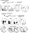
CD4 cells display different functional properties in the DLN than the lung. (A) BALB/c mice were infected with 500 EIU, and DLN and lung cells were isolated at various times postinfection to analyze the endogenous response. (B) Number of cells secreting IFN-γ per 1 × 10⁵ total input cells incubated with class II-restricted peptides HA126-138 and NP216-230. Cells were also incubated with the class I-restricted NP147-160 as a positive control. (C) At day 7 postinfection, DLN and lung cells were isolated and incubated with HA126-138-pulsed A20 cells, influenza virus-infected A20 cells, or anti-CD3 for 3 days, with [3H]thymidine added for the last 18 h. Shown is the average fold expansion over that for the medium control for three separate experiments. (D) DLN and lung cell preparations isolated at day 8 were incubated with antibodies to CD4, CD8, and GrB and subjected to flow cytometric analysis. A representative histogram of GrB expression gated on CD4 or CD8 cell populations is shown. (E) Total CD4 or CD8 T cell numbers (left axis) in the lungs of influenza virus-infected mice were calculated by multiplying total cell numbers by percent CD4- or CD8-positive cells analyzed by flow cytometry and the mean fluorescence intensity (MFI; right axis) of GrB expression in individual mice. *, P = 0.0001 by unpaired Student's t test. (F) HNT Thy1.1 CD4 cells (1 × 10⁶ to 2 × 10⁶) were adoptively transferred into healthy BALB/c mice, and the mice were subsequently infected with 500 EIU PR8. At 7 days postinfection, DLN, spleen, and lung cell populations were isolated as described in Materials and Methods and incubated with antibodies to CD4 and Thy1.1 to identify HNT peptide-specific CD4 cells. (G) Percentage of HNT cells in each organ at day 7 p.i. from a representative experiment. (H) Average percentage ± SD of CD4 Thy1.1 cells over time. (I) CD4 Thy1.1+ cells gated in panel G were analyzed for expression of GrB directly ex vivo. The gate depicts cells that are positive for expression based on the isotype control. (J) DLN, spleen, and lung cells isolated as in panel F were further restimulated with HA126-138 for 4 h in vitro and analyzed for the ability to produce IL-2 and IFN-γ by flow cytometry. Histograms show cells that were gated on CD4 and Thy1.1, as in panel G. The gate depicts cells that are positive for cytokine expression based on staining in the medium controls. Samples were run on a BD FACScaliber apparatus, and data were analyzed using Flow Jo software. (K) Average percentage ± SD of cytokine-secreting cells (left axis) or GrB MFI (right axis) of CD4/Thy1.1+ cells in various organs at 7 days postinfection in three separate experiments.
Fig 3
Fig 3
Lethal influenza virus infection induces class II expression on lung epithelial cells. Mice were given HNT Thy1.1+ effectors and subsequently infected with 5,000 EIU PR8. Five days later, mice were sacrificed and lungs were frozen as described in Materials and Methods. Shown is immunofluorescent analysis of frozen lung sections from uninfected mice (B), mice infected with 5,000 EIU PR8 (A and C), or infected mice given HNT Thy1.1+ CD4 cells (D) incubated with antibodies against class II (red) or Thy1.1 (green). (D) Staining of HNT-specific CD4 cells (green) in proximity to epithelial cells expressing class II (red). Magnification, ×200.
Fig 4
Fig 4
Lung resident CD4 cells express granzymes and perforin and lyse peptide-pulsed targets. (A and B) Mice were infected with 500 EIU, and at 8 days postinfection, CD4 cells were isolated from the DLN or lung as described in the text. The non-CD4 cells were used as a control that contained CD8 T cells. Shown is the relative quantity of T-bet and Eomes (A) or GrA, GrB, and perforin (B) mRNA by real-time PCR using CD4 cells from the DLN as the reference sample. Lung CD4 cells express significantly higher levels of T-bet than Eomes (P = 0.006 by Student's unpaired t test). Shown is an average from three separate cell isolations. (C) Mice were infected and cells were isolated as described for panels A and B and assayed for cytotoxicity against A20 cells pulsed with influenza virus-specific class I peptides (NP147-156 and HA518-526) or class II peptides (HA126-138, NP216-230, and NP146-160) (left) or A20 cells pulsed with class II peptides or infected with influenza virus (right). This experiment was repeated, with similar results. HNT Thy1.1 cells (1 × 10⁶) were adoptively transferred to BALB/c mice that were then infected with 500 EIU PR8. E:T, effector-to-target cell ratio. (D and E) Total CD4 cells were isolated by magnetic beads which contain endogenous and TCR Tg CD4 cells (D) or Thy1.1+ cells, isolated by FACS, which are purified TCR Tg cells (E), from the DLN, spleen (Spl), and lung and incubated with HNT-pulsed A20 target cells in a 4-h cytotoxicity assay. Shown is the percent lysis of each population against peptide-pulsed (closed symbols) or unpulsed (open symbols) target cells. These experiments were repeated once (E) or twice (D) with similar results.
Fig 5
Fig 5
IFN-γ is not required for the multifunctional CD4 T cell response to influenza virus infection. (A) BALB/c (WT) or BALB/c mice deficient in IFN-γ (γ−/−) were infected with 500 EIU and their weights were monitored over time. (B) Mice were bled at the indicated time points and analyzed for total influenza virus-specific IgG (left) or influenza virus-specific IgG1 or IgG2a (right) at day 25 p.i. by enzyme-linked immunosorbent assay. Shown is the endpoint titer. P values were determined by Student's t test: *, P = 0.028; †, P = 0.002. (C) Cells from the DLN of WT and IFN-γ−/− mice were restimulated in vitro with various influenza virus-specific peptides for 72 h, and [3H]thymidine incorporation was measured as an indication of proliferation. Shown is the stimulation index calculated as the fold increase over cells incubated in medium alone of three separate experiments. (D) CD4 cells were isolated from the lungs of WT (closed symbols) or IFN-γ−/− (open symbols) influenza virus-infected mice at various time points and incubated with HA or NP peptides in an ELISpot assay for IFN-γ (closed symbols) or IL-17 (open symbols). Shown is the number of cytokine-secreting spots calculated for 1 × 10⁶ CD4+ cells. (E) CD4 cells were isolated from the lungs of WT (closed symbols) or IFN-γ−/− (open symbols) influenza virus-infected mice at various time points and incubated with PMA and ionomycin. The percentage of cytokine-positive cells after gating on activated (CD4+/CD43+) cells is shown over time for one representative experiment. (F) Shown is the average ± SD of percent cytokine-positive cells in WT and IFN-γ−/− CD4 cells restimulated with PMA and ionomycin at day 10 p.i. from three separate experiments. *, statistically significant difference in percent cytokine-positive cells between WT and IFN-γ−/− cells (P < 0.005).
Fig 6
Fig 6
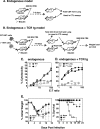
IFN-γ is dispensable for CTL activity but is necessary for complete protection from weight loss. (A) Schematic of endogenous CD4 effector generation for analysis of CTL activity. WT or IFN-γ−/− mice were infected with 500 EIU, and at day 9, mice were sacrificed and total CD4 cells were isolated for CTL assays (C). (B) Schematic of endogenous plus TCR Tg CD4 effector generation and transfer for CTL analysis (D) and protection studies. WT or IFN-γ−/− HNT cells are first transferred to WT or IFN-γ−/− mice before infection with 500 EIU. Total CD4 cells are then isolated as described at day 7 and used in CTL assays (D) or protection studies (E). (C) Total CD4 cells from the lung were isolated as described and assayed for cytotoxicity against A20 target cells pulsed with a mixture of class II peptides (closed symbols). Unpulsed A20 cells (open symbols) served as controls for antigen-specific killing. (D and E) Total CD4 cells were isolated from the lungs of mice receiving TCR Tg cells and assayed for cytotoxicity against HNT-pulsed A20 target cells (D) or transferred to healthy BALB/c mice that were then infected with 5,000 EIU PR8 and monitored for weight loss and survival (E). Shown is percent initial weight (left) and survival (right) over time from 10 individual mice. †, statistically significant difference in percent initial weight at day 9 (P = 0.002) by Student's t test; *, P = 0.06 by log-rank test.
Fig 7
Fig 7
Perforin is required for cytolytic activity by CD4 T cells in vivo and may be important for faster recovery from lethal influenza virus infection. (A) WT or Pfn−/− HNT cells were adoptively transferred to BALB/c Thy1.1 mice that were then infected with 500 EIU PR8. Total lung cells were isolated and incubated with antibodies to CD4 and Thy1.2 to detect adoptively transferred HNT cells. (B) Relative level of GrB expressed by CD4+ Thy1.2+ WT or Pfn−/− HNT cells after gating on the cells shown in panel A. (C) Total CD4 cells were isolated from the lung using anti-CD4-conjugated magnetic beads and assayed for cytotoxicity against HNT peptide-pulsed targets. Closed symbols denote effectors incubated with HNT peptide-pulsed targets, and open symbols denote effectors incubated with HNT peptide-pulsed targets in the presence of antibodies to FasL. (D) Schematic of transfer protocol for weight loss and survival studies. WT or Pfn−/− HNT cells were adoptively transferred to BALB/c mice that were then infected with 500 EIU PR8. At 7 days p.i., total CD4 cells were isolated from the lung and DLN and transferred to healthy BALB/c or JhD mice that were then infected with 5,000 EIU PR8 and monitored for weight loss and survival. (E) Percent initial weight over time for 10 BALB/c mice per group. (F) Total CD4 cells were isolated as described and transferred to JhD mice deficient in B cells. Percent initial weight (left) and survival (right) over time are shown for 10 mice per group. *, a statistically significant difference in percent initial weight at day 9 (P = 0.002) by Student's t test.

References

    1. Appay V. 2004. The physiological role of cytotoxic CD4(+) T-cells: the holy grail? Clin. Exp. Immunol. 138:10–13 - PMC - PubMed
    1. Appay V, et al. 2002. Characterization of CD4(+) CTLs ex vivo. J. Immunol. 168:5954–5958 - PubMed
    1. Baumgarth N, Kelso A. 1996. In vivo blockade of gamma interferon affects the influenza virus-induced humoral and the local cellular immune response in lung tissue. J. Virol. 70:4411–4418 - PMC - PubMed
    1. Boissonnas A, et al. 2010. Foxp3+ T cells induce perforin-dependent dendritic cell death in tumor-draining lymph nodes. Immunity 32:266–278 - PubMed
    1. Brien JD, Uhrlaub JL, Nikolich-Zugich J. 2008. West Nile virus-specific CD4 T cells exhibit direct antiviral cytokine secretion and cytotoxicity and are sufficient for antiviral protection. J. Immunol. 181:8568–8575 - PMC - PubMed

Publication types