. 2012 Sep;22(9):1374-89.
doi: 10.1038/cr.2012.57. Epub 2012 Apr 10.

Bidirectional regulation between WDR83 and its natural antisense transcript DHPS in gastric cancer

Affiliations

Bidirectional regulation between WDR83 and its natural antisense transcript DHPS in gastric cancer

Wen-Yu Su et al. Cell Res. 2012 Sep.

Abstract

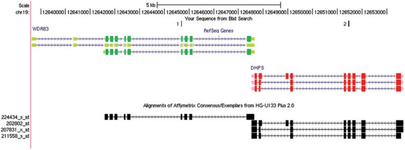
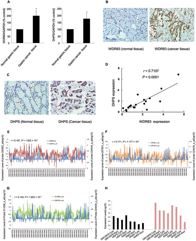
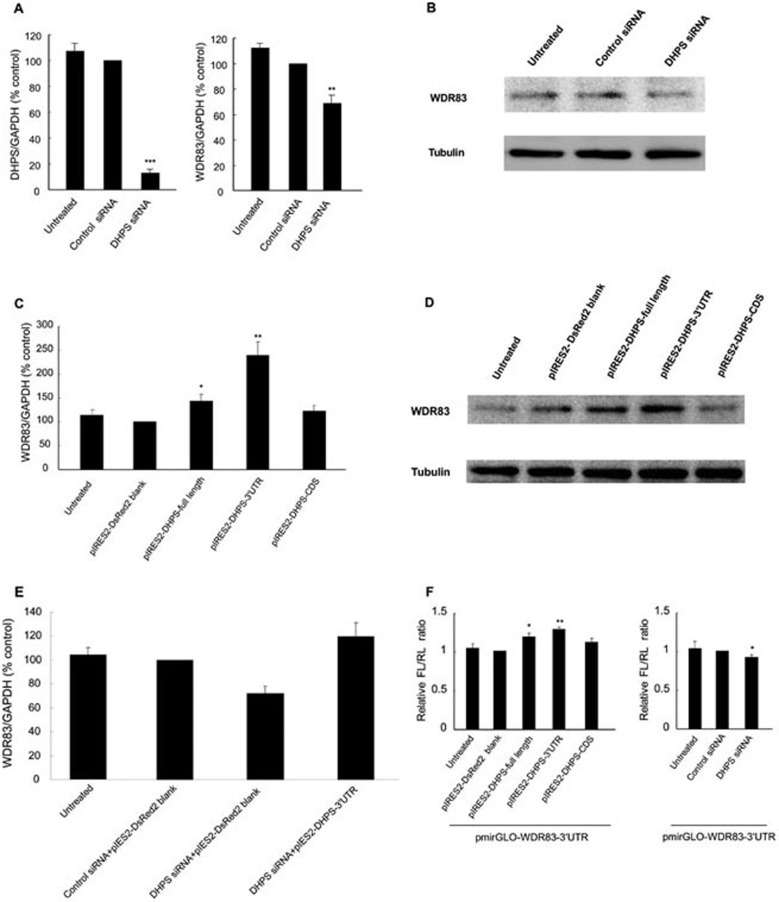
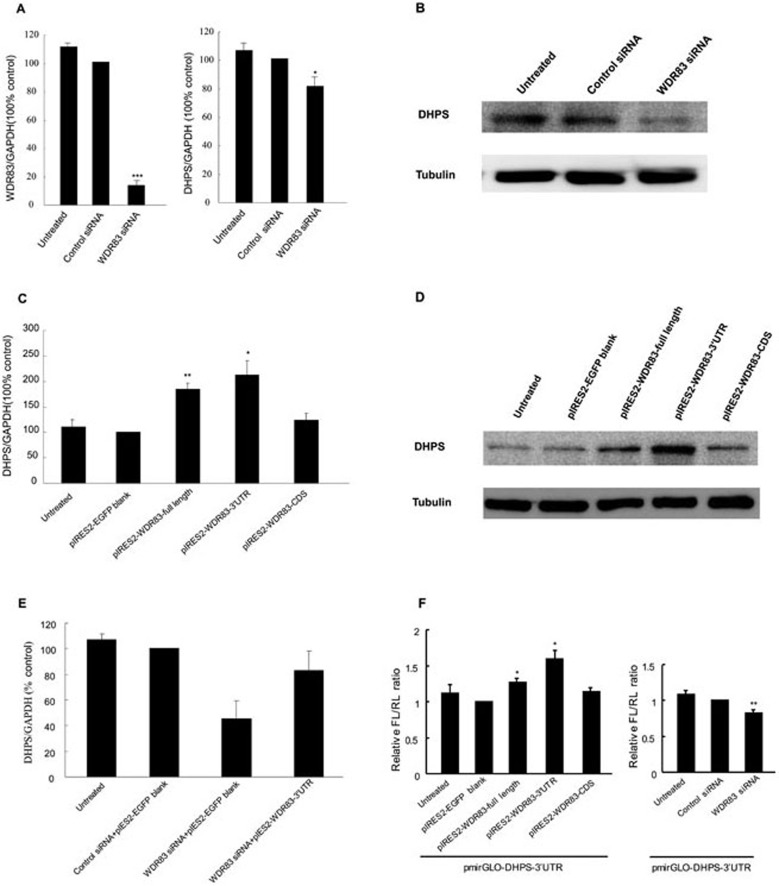
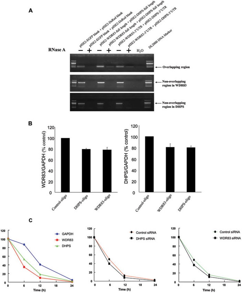
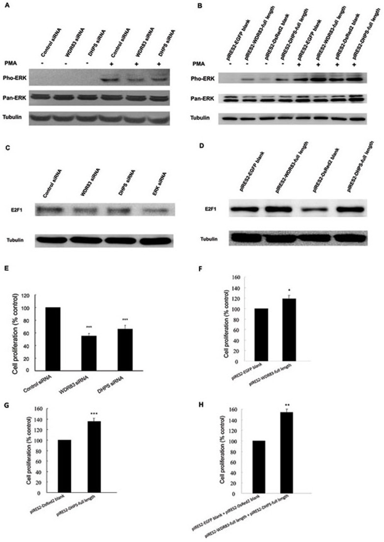
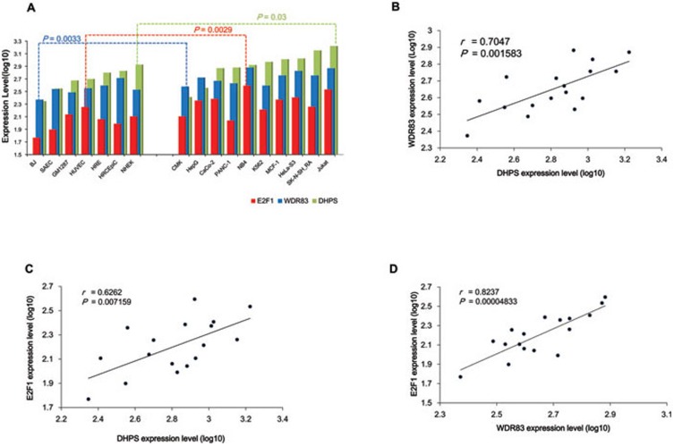
Natural antisense transcripts (NATs) exist ubiquitously in mammalian genomes and play roles in the regulation of gene expression. However, both the existence of bidirectional antisense RNA regulation and the possibility of protein-coding genes that function as antisense RNAs remain speculative. Here, we found that the protein-coding gene, deoxyhypusine synthase (DHPS), as the NAT of WDR83, concordantly regulated the expression of WDR83 mRNA and protein. Conversely, WDR83 also regulated DHPS by antisense pairing in a concordant manner. WDR83 and DHPS were capable of forming an RNA duplex at overlapping 3' untranslated regions and this duplex increased their mutual stability, which was required for the bidirectional regulation. As a pair of protein-coding cis-sense/antisense transcripts, WDR83 and DHPS were upregulated simultaneously and correlated positively in gastric cancer (GC), driving GC pathophysiology by promoting cell proliferation. Furthermore, the positive relationship between WDR83 and DHPS was also observed in other cancers. The bidirectional regulatory relationship between WDR83 and DHPS not only enriches our understanding of antisense regulation, but also provides a more complete understanding of their functions in tumor development.


Figures

Figure 1
Figure 1
Genomic organization of WDR83 and DHPS. DHPS and WDR83 are transcribed from opposite strands of the same region on chromosome 19. The coding regions and non-coding regions of WDR83 are marked with dark green and light green, respectively. The coding regions and noncoding regions of DHPS are marked with red and pink, respectively. The siRNA target sites of WDR83 (site 1) and DHPS (site 2) are located outside the overlapping regions to avoid off-target effects. The black boxes represent Affymetrix HG-U133 Plus2.0 array probe sets that correspond to WDR83 and DHPS. Arrows show the direction of transcription and blocks indicate exons.
Figure 2
Figure 2
Expression analysis and the subcellular location of WDR83 and DHPS in GC tissues and different cell lines. (A) WDR83 and DHPS mRNA levels were quantified by qPCR in 19 pairs of GC specimens and their matched normal tissues. WDR83 and DHPS expression were significantly increased in GC tissues compared with their normal tissues (P = 0.0198 and 0.0286, respectively, n = 19; *P< 0.05, **P< 0.01, ANOVA). (B, C) Representative expression of WDR83 and DHPS in GC specimens and their matched normal tissues as determined by IHC (magnification 200×); in GC tissues, high-level expression of WDR83 or DHPS was shown by strong staining. (D) Regression analysis of WDR83 and DHPS expression revealed that WDR83 and DHPS were positively correlated (r = 0.7107, P < 0.0001). The correlation coefficient (r) measured the reliability and the P-value measured the statistical significance of the correlation between the x and y variables. (E-G) Expression correlation analysis of WDR83 and DHPS in 504 normal tissues: (E) the expression of WDR83 (probe 224434_s_at, blue) and DHPS (probe 202802_at, red) exhibited a significantly positive correlation (r = 0.167, P< 0.00016); (F) the expression of WDR83 (probe 224434_s_at, blue) and DHPS (probe 207831_at, orange) also exhibited a significantly positive correlation (r = 0.171, P< 0.00012); (G) the expression of WDR83 (probe 224434_s_at, blue) and DHPS (probe 211558_s_at, green) exhibited concordant expression patterns (r = 0.140, P< 0.00017). (H) Subcellular location profiling of WDR83 and DHPS using the Affymetrix Tiling Array revealed that both genes were widely expressed in the cytoplasm and nucleus in different cell lines.
Figure 3
Figure 3
DHPS upregulated WDR83 mRNA and protein expression through the 3′UTRs. (A) Knockdown of DHPS by siRNAs in MGC803 cells reduced WDR83 mRNA levels by 31.4% (n ≥ 3, P = 0.0039). (B) Western blot analysis demonstrated that the siRNAs knockdown of DHPS decreased WDR83 protein expression. (C) Quantification of WDR83 mRNA levels after overexpression of three different plasmids in MGC803 cells. qPCR analysis indicated that overexpression of DHPS-full length or DHPS-3′UTR resulted in a significant increase in WDR83 mRNA levels (n ≥ 3, P = 0.0110 and 0.0080, respectively). (D) Western blot showing that WDR83 protein levels were significantly increased in MGC803 cells transfected with DHPS-full length or DHPS-3′UTR plasmids. (E) WDR83 mRNA levels were measured by qPCR in MGC803 cells where endogenous DHPS were first knocked down by siRNA for 24 h and then restored by exogenous expression of DHPS-3′UTR (pIRES2-DHPS-3′UTR) for further 24 h. (F) The luciferase reporter assay showed that the WDR83 3′UTR-containing construct induced a 24.4% increase in luciferase activity in cells that were cotransfected with the DHPS 3′UTR plasmid (P = 0.0020), and the WDR83 full-length construct also showed an increase in luciferase activity (P = 0.0310). The DHPS CDS-containing construct did not have an obvious effect on pmirGLO-WDR83-3′UTR (P = 0.7845). Meanwhile, silencing DHPS decreased luciferase activity (P = 0.0158). Relative firefly luciferase activity over renilla luciferase activity: FL/RL, n ≥ 3, ANOVA; *P< 0.05, **P< 0.01, ***P< 0.001.
Figure 4
Figure 4
WDR83 also upregulated DHPS mRNA and protein expression. (A) Knockdown of WDR83 by siRNAs in MGC803 cells resulted in an 18.7% decrease in DHPS mRNA expression (n ≥ 3, P = 0.0257). (B) Western blot analysis indicated that WDR83 downregulation also caused a decrease in DHPS protein expression. (C) Quantification of DHPS mRNA levels after overexpression of three different plasmids in MGC803 cells. qPCR analysis revealed that overexpression of only the transcripts containing the overlapping regions (WDR83-full length or WDR83-3′UTR) resulted in a significant increase in DHPS mRNA levels (n ≥ 3, P = 0.0014 and 0.0165, respectively). (D) Western blot analysis demonstrated that WDR83-full length or WDR83-3′UTR significantly increased DHPS protein levels in MGC803 cells. (E) Endogenous WDR83 was first knocked down by siRNA for 24 h and then restored by the exogenous expression of WDR83-3′UTR (pIRES2-WDR83-3′UTR) for further 24 h. Cells were harvested and the levels of DHPS mRNA were measured by qPCR. (F) The luciferase activity of pmirGLO-DHPS-3′UTR was increased significantly in cells that were cotransfected with the WDR83 3′UTR plasmid or the WDR83 full-length plasmid (P = 0.0481 and 0.0490, respectively), and the luciferase activity of pmirGLO-DHPS-3′UTR was markedly decreased in cells with knocked down WDR83 (P = 0.006; n ≥ 3, ANOVA; *P< 0.05, **P< 0.01, ***P< 0.001).
Figure 5
Figure 5
DHPS and WDR83 formed RNA duplex and increased stability of each other. (A) RPA was performed on RNA samples from MGC803 cells. The plasmids of pIRES2-WDR83-full length and pIRES2-DHPS-full length were cotransfected in the same reaction, and the plasmids of pIRES2-WDR83-3′UTR and pIRES2-DHPS-3′UTR were also cotransfected in the same reaction. Total RNA was extracted and purified, single-stranded RNA was digested with RNase A, and the remaining double-stranded RNA was subjected to RT-PCR to amplify the overlapping or non-overlapping regions of WDR83 and DHPS. (B) Blocking the WDR83/DHPS RNA-RNA interaction with 2′-O-methyl oligoribonucleotides inhibited the bidirectional regulation between them. (C) Stability of WDR83 and DHPS over time was measured by qPCR relative to time 0 after blocking new RNA synthesis with α-amanitin (25 mM). MGC803 cells were transfected with siRNA against WDR83 or DHPS or control siRNA for 24 h, and were then further exposed to 25 mM α-amanitin for 6, 12 and 24 h. Cells were harvested and the stability of the WDR83 and DHPS mRNA was analyzed by qPCR. 18S RNA, which was a product of RNA polymerase I and was unchanged after α-amanitin treatment, was used as the control.
Figure 6
Figure 6
WDR83 and DHPS were involved in the regulation of cell proliferation, and the downregulation of WDR83 or DHPS inhibited ERK1/2 activation and E2F1 expression in MGC803 cells. (A) The knockdown of either WDR83 or DHPS decreased PMA-stimulated ERK1/2 phosphorylation compared with negative controls in MGC803 cells. (B) Overexpression of WDR83 or DHPS in combination with PMA enhanced ERK1/2 phosphorylation. (C) The knockdown of WDR83, DHPS, or ERK1/2 resulted in a significant decrease in E2F1 protein levels compared with the control cells. (D) Overexpression of WDR83 or DHPS induced an increase in E2F1 protein levels. (E) Downregulation of WDR83 or DHPS significantly inhibited cell proliferation in MGC803 cells (P ≤ 0.0001 and P = 0.0006, respectively). (F, G) In contrast, the overexpression of WDR83 or DHPS enhanced cell proliferation (P = 0.0148 and P = 0.0003, respectively). (H) Interestingly, the joint overexpression of WDR83 and DHPS enhanced cell proliferation even further (P = 0.0037). Cell proliferation was determined by the CCK-8 assay (n ≥ 3, ANOVA; *P< 0.05, **P< 0.01, ***P< 0.001).
Figure 7
Figure 7
Positive correlations between WDR83, DHPS, and E2F1 in other cancers. The expressions of WDR83, DHPS, and E2F1 were analyzed in normal (n = 7) and cancer (n = 10) cell lines. Transcriptional profiles were derived from the ENCODE Transcriptome Project with the Affymetrix Human Exon 1.0 ST array. (A) The expression of WDR83, DHPS, and E2F1 was increased significantly in cancer cells compared with normal cells (P = 0.0033, 0.030, and 0.0029, respectively). (B-D) The expression of WDR83, E2F1, and DHPS exhibited positive correlation in multiple cell lines (P = 0.0016, 0.0071, and 0.00005, respectively).

Similar articles

Cited by

References

    1. Katayama S, Tomaru Y, Kasukawa T, et al. Antisense transcription in the mammalian transcriptome. Science. 2005;309:1564–1566. - PubMed
    1. Werner A, Sayer JA. Naturally occurring antisense RNA: function and mechanisms of action. Curr Opin Nephrol Hypertens. 2009;18:343–349. - PubMed
    1. Zhang Y, Liu XS, Liu QR, Wei L. Genome-wide in silico identification and analysis of cis natural antisense transcripts (cis-NATs) in ten species. Nucleic Acids Res. 2006;34:3465–3475. - PMC - PubMed
    1. Li JT, Zhang Y, Kong L, Liu QR, Wei L. Trans-natural antisense transcripts including noncoding RNAs in 10 species: implications for expression regulation. Nucleic Acids Res. 2008;36:4833–4844. - PMC - PubMed
    1. Beiter T, Reich E, Williams RW, Simon P. Antisense transcription: a critical look in both directions. Cell Mol Life Sci. 2009;66:94–112. - PMC - PubMed

Publication types

MeSH terms

Substances