

In vivo diagnosis of melanoma and nonmelanoma skin cancer using oblique incidence diffuse reflectance spectrometry
- PMID: 22491533
- PMCID: PMC3367032
- DOI: 10.1158/0008-5472.CAN-11-4027
In vivo diagnosis of melanoma and nonmelanoma skin cancer using oblique incidence diffuse reflectance spectrometry
Abstract
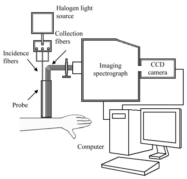
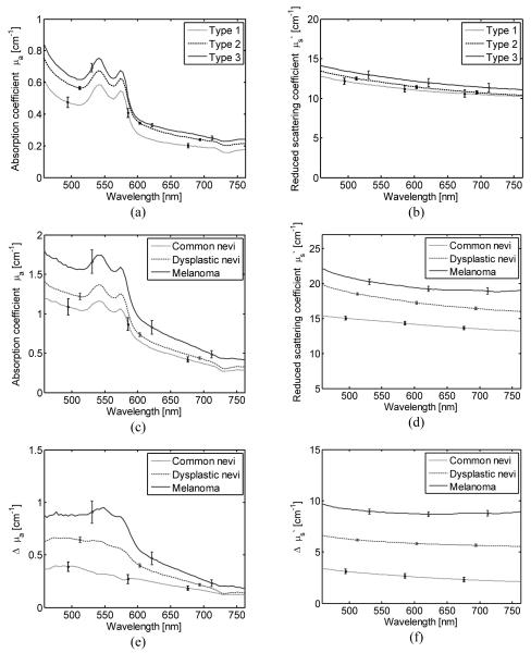
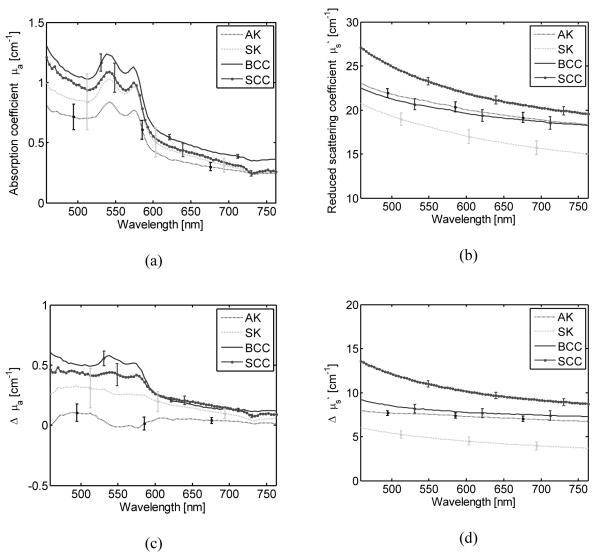
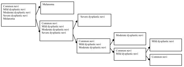
Early detection and treatment of skin cancer can significantly improve patient outcome. However, present standards for diagnosis require biopsy and histopathologic examinations that are relatively invasive, expensive, and difficult for patients with many early-stage lesions. Here, we show an oblique incidence diffuse reflectance spectroscopic (OIDRS) system that can be used for rapid skin cancer detection in vivo. This system was tested under clinical conditions by obtaining spectra from pigmented and nonpigmented skin lesions, including melanomas, differently staged dysplastic nevi, and common nevi that were validated by standard pathohistologic criteria. For diagnosis of pigmented melanoma, the data obtained achieved 90% sensitivity and specificity for a blinded test set. In a second analysis, we showed that this spectroscopy system can also differentiate nonpigmented basal cell or squamous cell carcinomas from noncancerous skin abnormalities, such as actinic keratoses and seborrheic keratoses, achieving 92% sensitivity and specificity. Taken together, our findings establish how OIDRS can be used to more rapidly and easily diagnose skin cancer in an accurate and automated manner in the clinic.
©2012 AACR
Figures
References
-
- American Cancer Society website Melanoma Skin Cancer. http://www.cancer.org.
-
- Cherpelis BS, Marcusen C, Lang PG. Prognostic Factors for Metastasis in Squamous Cell Carcinoma of the Skin. Dermatol Surg. 2002;28:268–73. - PubMed
-
- Takata M, Saida T. Early cancers of the skin: clinical, histopathological, and molecular characteristics. Int J Clin Oncol. 2005;10:391–97. - PubMed
-
- Zink D, Fischer AH, Nickerson JA. Nuclear structure in cancer cells. Nat Rev Cancer. 2004;4:677–87. - PubMed
Publication types
MeSH terms
Grants and funding
LinkOut - more resources
Full Text Sources
Medical
