Review
. 2012 May 1;4(5):a005777.
doi: 10.1101/cshperspect.a005777.

Synapses and Alzheimer's disease

Affiliations
Review

Synapses and Alzheimer's disease

Morgan Sheng et al. Cold Spring Harb Perspect Biol. .

Abstract

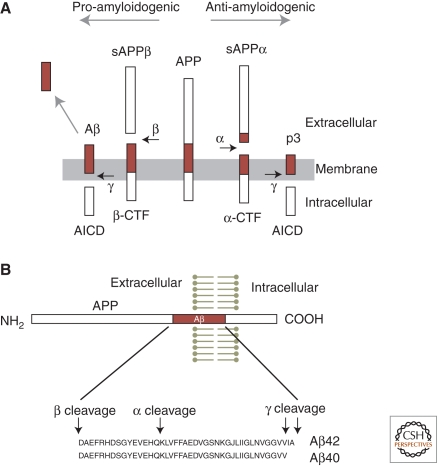
Alzheimer's disease (AD) is a major cause of dementia in the elderly. Pathologically, AD is characterized by the accumulation of insoluble aggregates of Aβ-peptides that are proteolytic cleavage products of the amyloid-β precursor protein ("plaques") and by insoluble filaments composed of hyperphosphorylated tau protein ("tangles"). Familial forms of AD often display increased production of Aβ peptides and/or altered activity of presenilins, the catalytic subunits of γ-secretase that produce Aβ peptides. Although the pathogenesis of AD remains unclear, recent studies have highlighted two major themes that are likely important. First, oligomeric Aβ species have strong detrimental effects on synapse function and structure, particularly on the postsynaptic side. Second, decreased presenilin function impairs synaptic transmission and promotes neurodegeneration. The mechanisms underlying these processes are beginning to be elucidated, and, although their relevance to AD remains debated, understanding these processes will likely allow new therapeutic avenues to AD.


Figures

Figure 1.
Figure 1.
APP processing and the formation of Aβ peptide. (A, middle) The full-length human amyloid precursor protein (APP), a single transmembrane protein with an intracellular carboxyl terminus. (Horizontal arrows) Specific protease cleavage sites. In the amyloidogenic pathway (to the left), sequential cleavage of APP by β-secretase and γ-secretase releases the soluble extracellular domain of APP (sAPPβ), Aβ peptide, and the intracellular carboxy-terminal domain of APP (AICD). Cleavage by α-secretase prevents formation of Aβ, instead producing sAPPα and p3 peptide. (CTF) Carboxy-terminal fragment of APP, before cleavage by γ-secretase. (B) Diagram of the APP polypeptide and sequence of Aβ40 and Aβ42 peptides, with secretase cleavage sites indicated.
Figure 2.
Figure 2.
Impaired spatial learning and memory in a transgenic mouse model of Alzheimer’s disease. Transgenic mice overexpressing human mutant APP and human mutant presenilin2 (PS2/APP mice) were tested in the acquisition of spatial memory in the Morris water maze at 6 mo of age. PS2/APP mice (n = 14) take significantly longer to reach a hidden platform during the 5 d of training (two sessions/day) than nontransgenic controls (NTG, n = 19). Repeated-measures ANOVA found a significant genotype (p < 0.001) and genotype × session interaction (p < 0.05). (Data kindly provided by William Meilandt, Tiffany Wu, and Kimberly Scearce-Levie [Genentech, Inc.].)
Figure 3.
Figure 3.
Effects of exogenous Aβ on dendritic spines and long-term potentiation in hippocampal slices. (A) Loss of dendritic spines induced by exposure to Aβ oligomers isolated from human AD brains. (Top) Image of an apical dendrite of a control CA1 hippocampal pyramidal neuron in an organotypic slice culture, showing the normal high density of dendritic spines. (Bottom) A similar neuron in a slice that has been exposed to ∼1 nm soluble Aβ oligomers derived from postmortem human AD brain. Prolonged exposure to Aβ oligomers from a variety of sources leads to loss of ∼50% of dendritic spines and of functional glutamatergic synapses. These images were acquired during the study described by Shankar et al. (2008) and are reprinted with permission from one of the authors. (B, top) Sustained long-term potentiation (LTP) is readily inducible by tetanic stimulation in untreated wild-type (WT) acute hippocampal slices (open symbols), but is blocked by exposure of the slice to soluble Aβ oligomers, especially at later time points (filled symbols). (Bottom) LTP is also inducible in caspase-3 knockout slices, but it is not suppressed by Aβ oligomers, indicating that caspase-3 is required for Aβ suppression of LTP. (These data were acquired by Kimberly Moore Olsen during the study described by Jo et al. [2011].)
Figure 4.
Figure 4.
Pathogenic hypotheses for synaptic and neuronal toxicity in Alzheimer’s disease. The specific hypotheses shown are not mutually exclusive, and, moreover, they likely “cross-talk” with each other. For instance, Aβ may induce tau hyperphosphorylation and aggregation, and presenilin mutations may cause lysosome and autophagy dysfunction (Pimplikar et al. 2010; Nixon and Yang 2011). Not all possible mechanisms of synaptic and neural toxicity are shown here (see text for additional examples).

References

    1. Ashe KH, Zahs KR 2010. Probing the biology of Alzheimer’s disease in mice. Neuron 66: 631–645 - PMC - PubMed
    1. Ballatore C, Lee VM, Trojanowski JQ 2007. Tau-mediated neurodegeneration in Alzheimer’s disease and related disorders. Nat Rev Neurosci 8: 663–672 - PubMed
    1. Bancher C, Braak H, Fischer P, Jellinger KA 1993. Neuropathological staging of Alzheimer lesions and intellectual status in Alzheimer’s and Parkinson’s disease patients. Neurosci Lett 162: 179–182 - PubMed
    1. Bettens K, Sleegers K, Van Broeckhoven C 2010. Current status on Alzheimer disease molecular genetics: From past, to present, to future. Hum Mol Genet 19: R4–R11 - PMC - PubMed
    1. Bezprozvanny I, Mattson MP 2008. Neuronal calcium mishandling and the pathogenesis of Alzheimer’s disease. Trends Neurosci 31: 454–463 - PMC - PubMed

Publication types

MeSH terms