

Fluoroscopy-guided retrograde core drilling and cancellous bone grafting in osteochondral defects of the talus
- PMID: 22491802
- PMCID: PMC3535023
- DOI: 10.1007/s00264-012-1530-9
Fluoroscopy-guided retrograde core drilling and cancellous bone grafting in osteochondral defects of the talus
Abstract
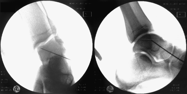
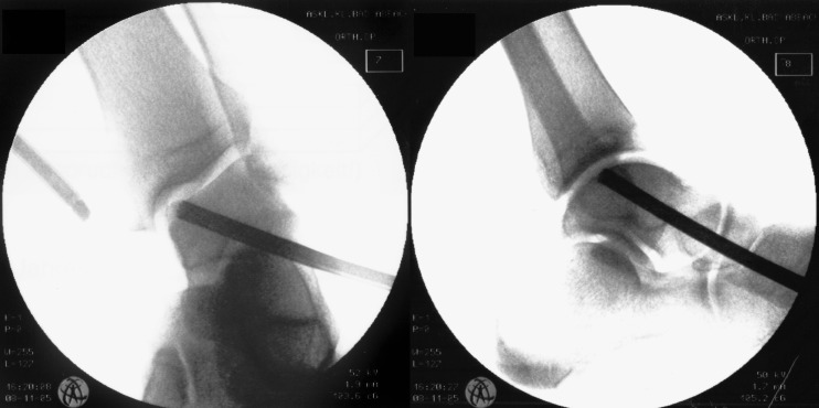
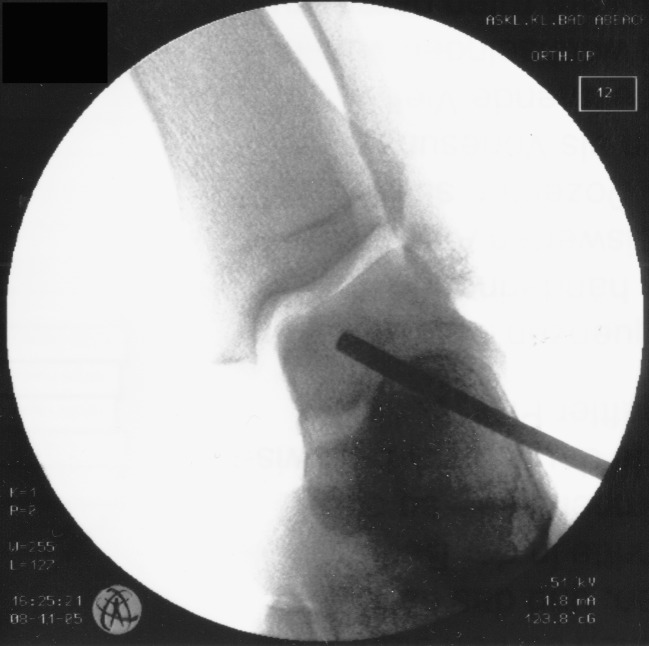
Purpose: In undetached osteochondral lesions (OCL) of the talus both revitalisation of the subchondral necrosis and cartilage preservation are essential. For these cases, we assess the results of minimally invasive retrograde core drilling and cancellous bone grafting.
Methods: Forty-one osteochondral lesions of the talus (12x grade I, 22x grade II and 7x grade III according to the Pritsch classification, defect sizes 7-14 mm) in 38 patients (mean age 33.2 years) treated by fluoroscopy-guided retrograde core drilling and autologous cancellous bone grafting were evaluated by clinical scores and MRI. The mean follow-up was 29.0 (±13) months.
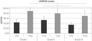
Results: The AOFAS score increased significantly from 47.3 (±15.3) to 80.8 (±18.6) points. Lesions with intact cartilage (grades I and II) had a tendency to superior results than grade III lesions (83.1 ± 17.3 vs. 69.4 ± 22.2 points, p = 0.07). First-line treatments and open distal tibial growth plates led to significantly better outcomes (each p < 0.05). Age, gender, BMI, time to follow-up, defect localisation or a traumatic origin did not influence the score results. On a visual analogue scale pain intensity reduced from 7.5 (±1.5) to 3.7 (±2.6) while subjective function increased from 4.6 (±2.0) to 8.2 (±2.3) (each p < 0.001). In MRI follow-ups, five of the 41 patients showed a complete bone remodelling. In two cases demarcation was detectable.
Conclusions: The technique reported is a highly effective therapeutic option in OCL of the talus with intact cartilage grades I and II. However, second-line treatments and grade III lesions with cracked cartilage surface can not be generally recommended for this procedure.
Figures

References
-
- Hangody L, Kish G, Modis L, Szerb I, Gaspar L, Dioszegi Z, Kendik Z. Mosaicplasty for the treatment of osteochondritis dissecans of the talus: two to seven year results in 36 patients. Foot Ankle Int. 2001;22:552–558. - PubMed
-
- Becher C, Thermann H. Results of microfracture in the treatment of articular cartilage defects of the talus. Foot Ankle Int. 2005;26:583–589. - PubMed
MeSH terms
LinkOut - more resources
Full Text Sources
Medical
Miscellaneous
