. 2012 May;17(3):331-5.
doi: 10.1007/s00776-012-0209-9. Epub 2012 Apr 11.

Neural mechanisms underlying deafferentation pain: a hypothesis from a neuroimaging perspective

Affiliations

Neural mechanisms underlying deafferentation pain: a hypothesis from a neuroimaging perspective

Takashi Hanakawa. J Orthop Sci. 2012 May.

Abstract

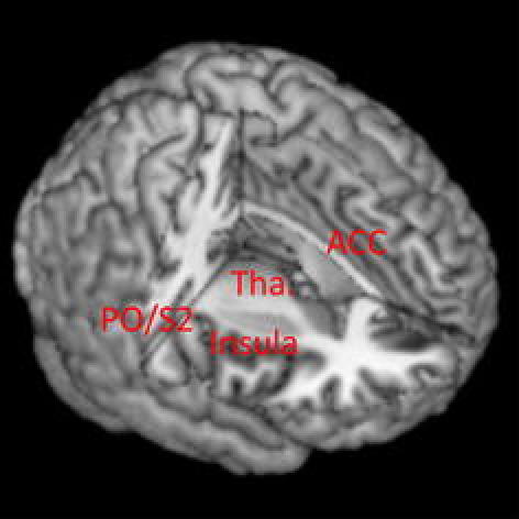
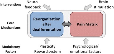
Deafferentation pain following nerve injury annoys patients, and its management is a challenge in clinical practice. Although the mechanisms underlying deafferentation pain remain poorly understood, progress in the development of multidimensional neuroimaging techniques is casting some light on these issues. Deafferentation pain likely results from reorganization of the nervous system after nerve injury via processes that interact with the substrates for pain perception (the pain matrix). Therapeutic effects of motor cortex stimulation on deafferentation pain suggest that the core mechanisms underlying deafferentation pain also interact with the motor system. Therefore, simultaneous neuroimaging and brain stimulation, an emerging neuroimaging technique, was developed to investigate complicated interactions among motor, somatosensory, and pain systems. In healthy participants, parts of the pain matrix (the anterior cingulate cortex, parietal operculum, and thalamus) show activity during both somatosensory stimulation and brain stimulation to the motor cortex. This finding indicates that motor, somatosensory, and pain systems communicate among each other via the neural network. A better understanding of the plastic mechanisms influencing such cross-talk among these systems will help develop therapeutic interventions using brain stimulation and neurofeedback.


Figures

Fig. 1
Fig. 1
Pain matrix. The anterior cingulate cortex (ACC), posterior insula, parietal operculum (PO) including the second somatosensory cortex (S2) and thalamus (Tha.) are considered to be the key structures for pain perception. The primary motor and somatosensory areas may be involved in the perception of nonthermal (deep) pain
Fig. 2
Fig. 2
Brain activities induced by deep somatosensory stimulation (blue) and transcranial magnetic stimulation to the primary motor cortex (red) in healthy volunteers. The overlap of the two is shown in green. The overlapping activity partially corresponds to the pain matrix, which is composed of the cingulate cortex, the insula/parietal operculum including the second somatosensory cortex, and the thalamus. Overlap is also observed in nonpain matrix areas, such as the primary motor cortex, the primary somatosensory cortex, the supplementary motor cortex, and the basal ganglia. The figure shows a new illustration based on data from Shitara et al. [20]
Fig. 3
Fig. 3
Hypothetical mechanisms of deafferentation pain. Deafferentation pain may result from an interaction between reorganization processes after deafferentation and their influences on the pain matrix (core mechanisms). These processes and perception of deafferentation pain are likely to be influenced by many factors, including plasticity, the reward system, and psychological/emotional factors. We need to look for therapeutic interventions (e.g., brain stimulation, neurofeedback) that can effectively modulate the activity of these core mechanisms, most likely represented in the form of a neural network

References

    1. Kooijman CM, Dijkstra PU, Geertzen JH, Elzinga A, Schans CP. Phantom pain and phantom sensations in upper limb amputees: an epidemiological study. Pain. 2000;87:33–41. doi: 10.1016/S0304-3959(00)00264-5. - DOI - PubMed
    1. Jensen TS, Krebs B, Nielsen J, Rasmussen P. Phantom limb, phantom pain and stump pain in amputees during the first 6 months following limb amputation. Pain. 1983;17:243–256. doi: 10.1016/0304-3959(83)90097-0. - DOI - PubMed
    1. Jensen TS, Krebs B, Nielsen J, Rasmussen P. Immediate and long-term phantom limb pain in amputees: incidence, clinical characteristics and relationship to pre-amputation limb pain. Pain. 1985;21:267–278. doi: 10.1016/0304-3959(85)90090-9. - DOI - PubMed
    1. Karl A, Birbaumer N, Lutzenberger W, Cohen LG, Flor H. Reorganization of motor and somatosensory cortex in upper extremity amputees with phantom limb pain. J Neurosci. 2001;15(21):3609–3618. - PMC - PubMed
    1. Mercier C, Reilly KT, Vargas CD, Aballea A, Sirigu A. Mapping phantom movement representations in the motor cortex of amputees. Brain. 2006;129(Pt 8):2202–2210. doi: 10.1093/brain/awl180. - DOI - PubMed

Publication types