. 2012 May;139(10):1831-41.
doi: 10.1242/dev.072850. Epub 2012 Apr 4.

Bcl11a is required for neuronal morphogenesis and sensory circuit formation in dorsal spinal cord development

Affiliations

Bcl11a is required for neuronal morphogenesis and sensory circuit formation in dorsal spinal cord development

Anita John et al. Development. 2012 May.

Abstract

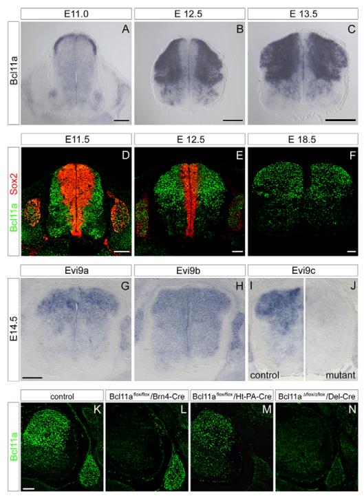
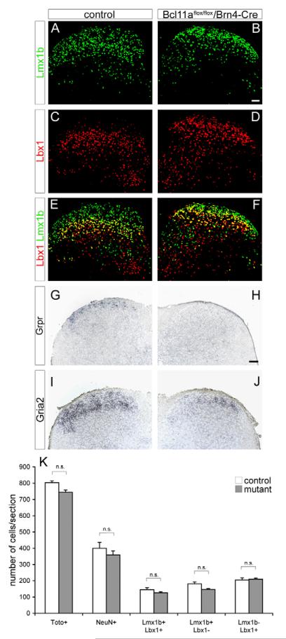
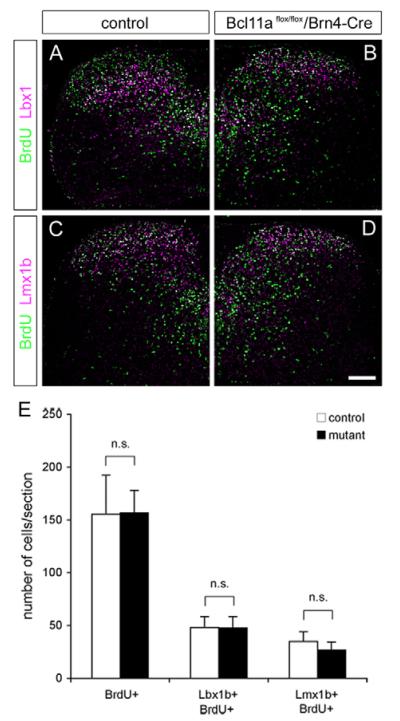
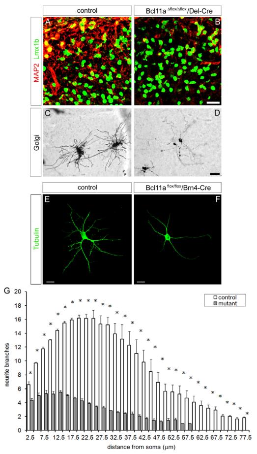
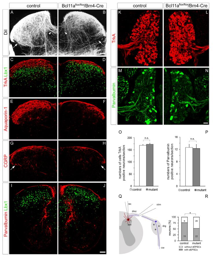
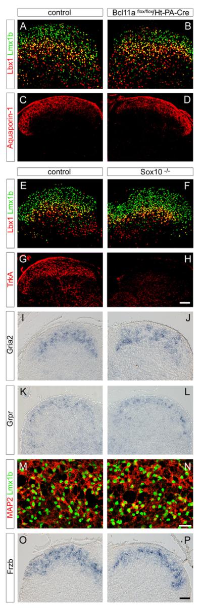
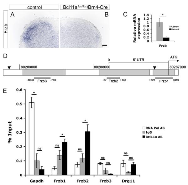
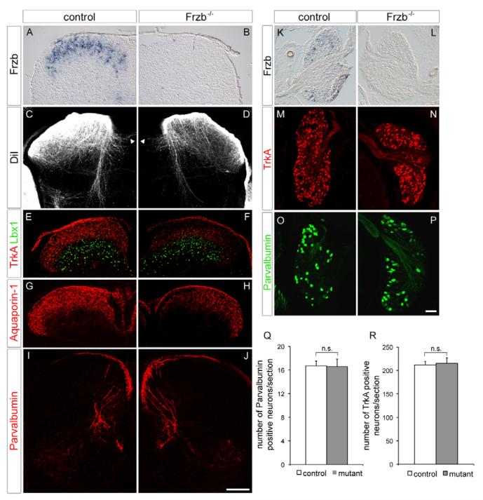
Dorsal spinal cord neurons receive and integrate somatosensory information provided by neurons located in dorsal root ganglia. Here we demonstrate that dorsal spinal neurons require the Krüppel-C(2)H(2) zinc-finger transcription factor Bcl11a for terminal differentiation and morphogenesis. The disrupted differentiation of dorsal spinal neurons observed in Bcl11a mutant mice interferes with their correct innervation by cutaneous sensory neurons. To understand the mechanism underlying the innervation deficit, we characterized changes in gene expression in the dorsal horn of Bcl11a mutants and identified dysregulated expression of the gene encoding secreted frizzled-related protein 3 (sFRP3, or Frzb). Frzb mutant mice show a deficit in the innervation of the spinal cord, suggesting that the dysregulated expression of Frzb can account in part for the phenotype of Bcl11a mutants. Thus, our genetic analysis of Bcl11a reveals essential functions of this transcription factor in neuronal morphogenesis and sensory wiring of the dorsal spinal cord and identifies Frzb, a component of the Wnt pathway, as a downstream acting molecule involved in this process.


Figures

Fig. 1
Fig. 1. Expression analysis and conditional mutation of Bcl11a in the spinal cord
(A-F) In situ hybridization (A-C) and immunohistological analysis (D-F) of Bcl11a expression in the embryonic spinal cord at different developmental stages using Bcl11a-specific probes and antibodies, respectively. The ventricular zone was visualized with antibodies against Sox2 (D,E). (G-J) In situ hybridizations using Evi9a (G), Evi9b (H) and Evi9c (I,J) specific probes on wild-type (G-I) or Bcl11a mutant (J) spinal cord sections of E14.5 embryos. (K-N) Immunohistological analysis of spinal cord and dorsal root ganglia (DRG) on transverse sections of control (K), Bcl11aflox/flox;Brn4-Cre (L), Bcl11aflox/flox;Ht-PA-Cre (M) and Bcl11aΔflox/Δflox;Del-Cre (N) mouse embryos at E15.5 using antibodies against Bcl11a. Scale bars: 100 μm.
Fig. 2
Fig. 2. Analysis of neuronal differentiation in the Bcl11a mutant dorsal spinal cord
(A-F) Immunohistological analysis on dorsal spinal cord sections from control (A,C,E) and Bcl11aflox/flox;Brn4-Cre (B,D,F) mice at E18.5 using antibodies against Lmx1b (A,B,E,F) or Lbx1 (C-F). (G-J) In situ hybridization analysis on dorsal spinal cord sections from control (G,I) and Bcl11a mutant (H,J) animals at E18.5, with probes specific for Grpr (G,H) and Gria2 (I,J). (K) Numbers of cells (Toto+), neurons (NeuN+) and of marker-defined neuron populations in the dorsal horn of Bcl11a mutant and control embryos. Mean±s.e.m. n.s., not significant. Scale bars: 50 μm.
Fig. 3
Fig. 3. Birthdating and tracing of Bcl11a mutant dorsal spinal neurons
(A-D) Immunohistochemical analysis of dorsal spinal cord on transverse sections of control (A,C) and Bcl11aflox/flox;Brn4-Cre (B,D) mice that received a single dose of BrdU at E11.5 and were sacrificed at E16.5 using antibodies against BrdU (A-D), Lbx1 (A,B) and Lmx1b (C,D). (E) Numbers of BrdU+, Lbx1+ BrdU+ and Lmx1b+ BrdU+ cells in the spinal cord of Bcl11a mutant and control embryos. Mean±s.e.m. n.s., not significant. Scale bar: 100 μm.
Fig. 4
Fig. 4. Dorsal spinal neurons require Bcl11a for morphogenesis
(A-D) Transverse sections of the superficial dorsal horn of control (A,C) and Del-Cre recombined Bcl11a mutant (B,D) mice. Note that in mutants, Lmx1b-expressing neurons are more densely packed (B). Neurite morphology of control (C) and Bcl11a mutant (D) neurons in the superficial dorsal horn at E18.5 was visualized by Golgi staining. (E,F) Primary neuron cultures derived from dorsal spinal cord tissues of E18.5 control (E) and Bcl11a mutant (F) animals stained with anti-tubulin antibodies. (G) Sholl analysis of neurite length and arborization. Mean±s.e.m. *, P<0.05 (Student’s t-test). Scale bars: 20 μm in B; 30 μm in D; 40 μm in E,F.
Fig. 5
Fig. 5. Sensory axon projections in the Bcl11a mutant dorsal spinal horn
(A-J) Transverse sections of the dorsal horn of control (A,C,E,G,I) and Brn4-Cre recombined Bcl11a mutant (B,D,F,H,J) mice at E16.5 (A,B) and E18.5 (C-J). Sensory axons were traced with DiI (A,B). Axons crossing the midline are marked (arrowheads in A,B). Residual fibers are disorganized (arrows in A,B). A similar phenotype is detectable with anti-CGRP staining (arrows in G,H). (C-J) Immunohistological analysis of sensory axons projecting into the spinal cord with antibodies against TrkA (C,D), aquaporin 1 (E,F), CGRP (G,H) and parvalbumin (I,J). (K-P) Immunohistological detection (K-N) and quantification (O,P) of TrkA-positive (K,L,O) and parvalbumin-positive (M,N,P) sensory neurons in DRG. Mean±s.e.m. n.s., not significant. (Q,R) Electrophysiological analysis of evoked excitatory postsynaptic currents (eEPSCs) from dorsal horn neurons of control and Brn4-Cre recombined Bcl11a mutants at E18.5. *, P<0.05 (Fisher’s exact test). rec, recording pipette; stim, stimulation pipette; dsn, dorsal spinal neuron; drez, dorsal root entry zone; drg, dorsal root ganglion; csa, central sensory axon. Scale bars: 50 μm.
Fig. 6
Fig. 6. Analysis of dorsal spinal cords of Ht-PA-Cre recombined Bcl11a mutants and of Sox10 mutants
(A-P) Transverse sections of the dorsal horn from control (A,C,E,G,I,K,M,O), Ht-PA-Cre recombined Bcl11a mutant (B,D) and Sox10 mutant (F,H,J,L,N,P) mice at E18.5, stained with antibodies against Lmx1b (A,B,E,F,M,N), Lbx1 (A,B,E,F), aquaporin 1 (C,D), TrkA (G,H) and MAP2 (M,N), or hybridized with probes against Gria2 (I,J), Grpr (K,L) and Frzb (O,P). (M,N) High magnifications of the superficial zones of dorsal horns from controls (M) and Sox10 mutants (N). Scale bars: 50 μm in H,P; 20 μm in N.
Fig. 7
Fig. 7. Analysis of target gene expression in Bcl11a mutant dorsal spinal cord
(A,B) In situ hybridization analysis of Frzb mRNA expression in the dorsal horn of controls (A) and Brn4-Cre recombined Bcl11a mutants (B) at E14.5. Scale bar: 50 μm. (C) Determination of Frzb mRNA expression levels of control and mutant dorsal spinal cords by qRT-PCR at E14.5. Mean±s.e.m. *, P<0.05. (D) The Frzb promoter region indicating candidate regulatory regions (gray) as predicted by Ensembl Genome Browser and showing amplified fragments Frzb1-3 used in ChIP analysis. The 5′UTR and ATG are indicated. Black arrowheads mark the boundaries of a Frzb promoter fragment used previously to drive transgenic Frzb expression in mice (Tylzanowski et al., 2004). (E) ChIP assays on wild-type dorsal spinal cord tissue at E14.5 employing specific antibodies against Bcl11a or RNA polymerase II, and IgG as control. Binding of Bcl11a or RNA polymerase II to DNA was determined by qPCR using specific primer pairs for the Frzb promoter regions as indicated in D (region 1, +849 to +623; region 2, +138 to −27; region 3, −786 to −1008, relative to the transcription start site) as well as specific primer pairs amplifying the Gapdh and Drg11 promoters. Pairwise comparison of ChIP data employing specific antibodies (RNA Pol AB, Bcl11a AB) versus IgG negative control was used for statistical analysis. Significantly more Frzb promoter DNA of regions 1 and 2 was recovered with the Bcl11a antibody than in controls Mean±s.e.m.; n=3. *, P<0.05 (Student’s t-test); ns, not significant.
Fig. 8
Fig. 8. Sensory axon projections in the Frzb mutant dorsal spinal horn
(A-J) Transverse sections of the dorsal horn of control (A,C,E,G,I) and Frzb mutant (B,D,F,H,J) mice at E16.5 (A-D) and E18.5 (E-J) hybridized with probes specific for Frzb (A,B) or stained with antibodies against TrkA (E,F), aquaporin 1 (G,H) or parvalbumin (I,J). Sensory axons entering the dorsal horn were traced with DiI (C,D). Axons crossing the midline are marked by arrowheads (C,D). (K-P) transverse sections of DRG of controls (K,M,O) and Frzb mutants (L,N,P) hybridized with Frzb-specific probes (K,L) or stained with antibodies against TrkA (M,N) and parvalbumin (O,P). (Q,R) Quantitative analysis of sensory neurons in DRG. Mean±s.e.m. n.s., not significant. Scale bars: 100 μm in J; 50 μm in P.

References

    1. Arlotta P, Molyneaux BJ, Chen J, Inoue J, Kominami R, Macklis JD. Neuronal subtype-specific genes that control corticospinal motor neuron development in vivo. Neuron. 2005;45:207–221. - PubMed
    1. Arlotta P, Molyneaux BJ, Jabaudon D, Yoshida Y, Macklis JD. Ctip2 controls the differentiation of medium spiny neurons and the establishment of the cellular architecture of the striatum. J. Neurosci. 2008;28:622–632. - PMC - PubMed
    1. Avram D, Fields A, Pretty On Top K, Nevrivy DJ, Ishmael JE, Leid M. Isolation of a novel family of C(2)H(2) zinc finger proteins implicated in transcriptional repression mediated by chicken ovalbumin upstream promoter transcription factor (COUP-TF) orphan nuclear receptors. J. Biol. Chem. 2000;275:10315–10322. - PMC - PubMed
    1. Avram D, Fields A, Senawong T, Topark-Ngarm A, Leid M. COUP-TF (chicken ovalbumin upstream promoter transcription factor)-interacting protein 1 (CTIP1) is a sequence-specific DNA binding protein. Biochem. J. 2002;368:555–563. - PMC - PubMed
    1. Baccei ML, Fitzgerald M. Development of GABAergic and glycinergic transmission in the neonatal rat dorsal horn. J. Neurosci. 2004;24:4749–4757. - PMC - PubMed

Publication types

MeSH terms