Comparative Study
. 2012 Apr 4;32(14):4705-15.
doi: 10.1523/JNEUROSCI.0169-12.2012.

Age-dependent rescue by simvastatin of Alzheimer's disease cerebrovascular and memory deficits

Affiliations
Comparative Study

Age-dependent rescue by simvastatin of Alzheimer's disease cerebrovascular and memory deficits

Xin-Kang Tong et al. J Neurosci. .

Erratum in

  • J Neurosci. 2012 May 30;32(22):7766. Rosa-Neto, Pedro [added]

Abstract

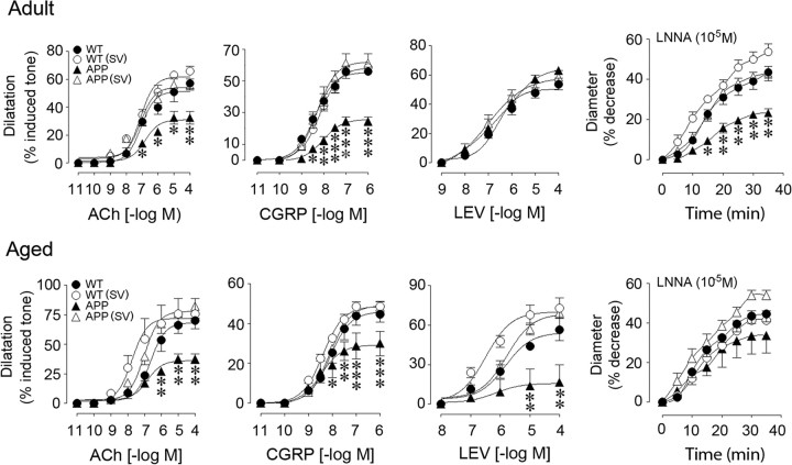
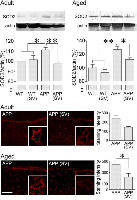
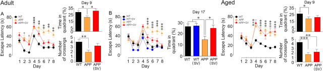
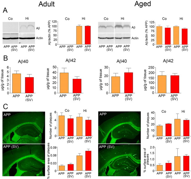
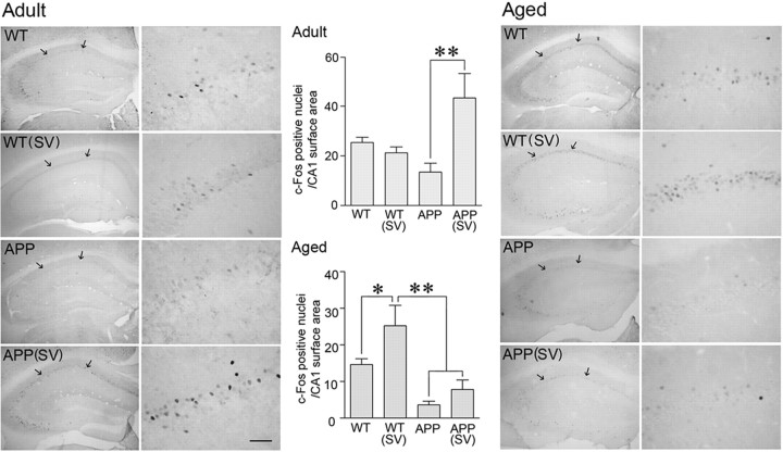
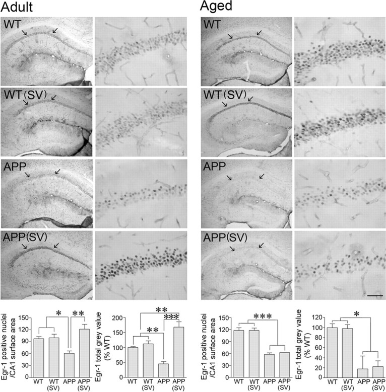
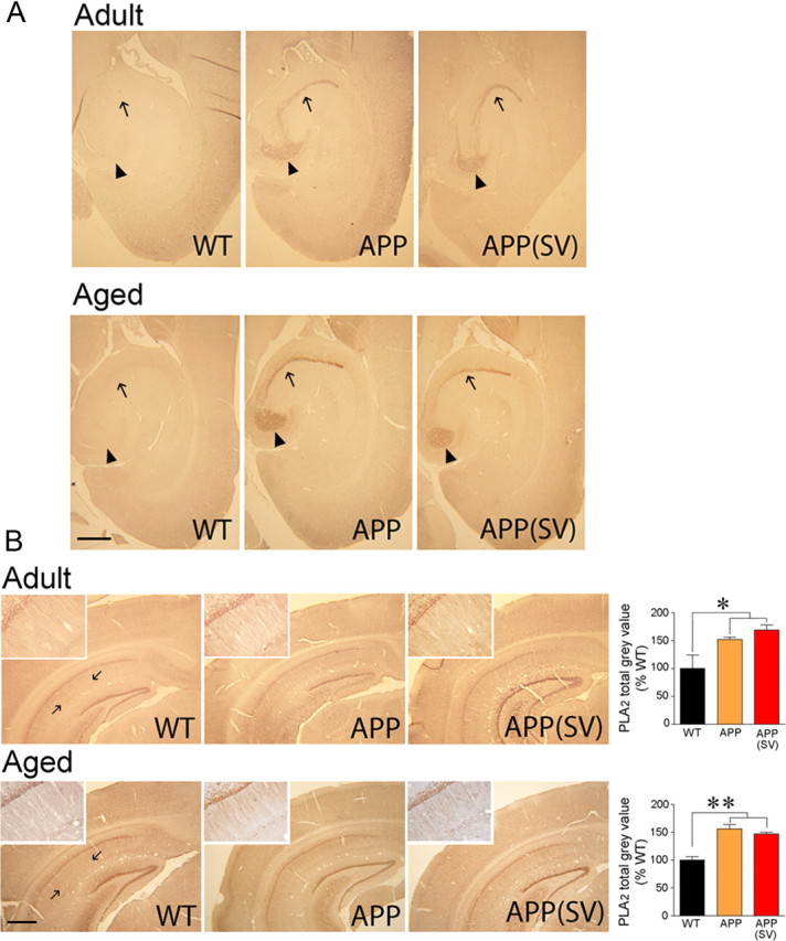
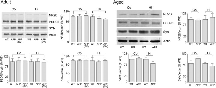
Alzheimer's disease (AD) is now established as a progressive compromise not only of the neurons but also of the cerebral vasculature. Increasing evidence also indicates that cerebrovascular dysfunction may be a key or an aggravating pathogenic factor in AD, emphasizing the importance to properly control this deficit when aiming for effective therapy. Here, we report that simvastatin (3-6 months, 40 mg/kg/d) completely rescued cerebrovascular reactivity, basal endothelial nitric oxide synthesis, and activity-induced neurometabolic and neurovascular coupling in adult (6 months) and aged (12 months) transgenic mice overexpressing the Swedish and Indiana mutations of the human amyloid precursor protein (AD mice). Remarkably, simvastatin fully restored short- and long-term memory in adult, but not in aged AD mice. These beneficial effects occurred without any decreasing effect of simvastatin on brain amyloid-β (Aβ) levels or plaque load. However, in AD mice with recovered memory, protein levels of the learning- and memory-related immediate early genes c-Fos and Egr-1 were normalized or upregulated in hippocampal CA1 neurons, indicative of restored neuronal function. In contrast, the levels of phospholipase A2, enkephalin, PSD-95, synaptophysin, or glutamate NMDA receptor subunit type 2B were either unaltered in AD mice or unaffected by treatment. These findings disclose new sites of action for statins against Aβ-induced neuronal and cerebrovascular deficits that could be predictive of therapeutic benefit in AD patients. They further indicate that simvastatin and, possibly, other brain penetrant statins bear high therapeutic promise in early AD and in patients with vascular diseases who are at risk of developing AD.


Figures

Figure 1.
Figure 1.
Simvastatin (SV) rescued cerebrovascular reactivity in adult and aged APP mice. The impaired dilatations to ACh and CGRP in APP mice (▴) compared with WT controls (●) were normalized by SV treatment (▵), which had no effect in WT mice (○). Dilatation to the KATP channel opener levcromakalim (LEV) was unaltered in adult APP mice, but was reduced in aged APP mice and completely restored by SV. Contractile responses to NOS inhibition (l-NNA, 10−5 m) were significantly reduced only in adult APP mice, and were normalized by SV. *p < 0.05; **p < 0.01; ***p < 0.001 for APP vs WT or APP (SV) mice. ANOVA and Newman–Keuls test, n = 4/group.
Figure 2.
Figure 2.
Simvastatin (SV) decreased oxidative stress in brain vessels and pial membranes from APP mice. In both adult and aged APP mice, SV normalized the increased SOD2 protein levels in cerebral arteries measured by Western blot (top) (n = 5–13 mice/group in adult and n = 4 mice/group in aged mice). SV also reduced the staining intensity of the O2 marker hydroethidine in the pial membrane (bottom) and intracortical blood vessels (not quantified). *p < 0.05; **p < 0.01 (n = 4–5 mice /group at both ages). Scale bar, 30 μm.
Figure 3.
Figure 3.
Simvastatin (SV) normalized neurovascular and neurometabolic coupling in APP mice. A, SV normalized the impaired CBF response to whisker stimulation (shaded area) in adult and aged APP mice relative to WT controls. Curves represent 1 s average of the evoked CBF, expressed as a percentage change from 30 s prestimulus baseline [black, WT; blue, WT (SV), orange, APP; red, APP (SV)]. *p < 0.05; **p < 0.001. B, The impaired increase in glucose uptake after whisker stimulation in aged APP mice was restored by SV. The percentage specific activation was calculated from the [18F]FDG SUV in the activated contralateral and analogous ipsilateral somatosensory cortex (as indicated by arrows on the merged images). Average PET images were overlaid over average anatomical MRI images obtained from similarly aged WT and APP mice. *p < 0.05 compared with WT and APP(SV) mice. ANOVA and Newman–Keuls test, n = 4–7 per group.
Figure 4.
Figure 4.
Simvastatin (SV) rescued learning and memory in adult but not in aged APP mice. APP mice of both ages (orange triangles) displayed longer latencies to reach the hidden platform (days 4–8), as well as decreased time and number of platform crossings in the target quadrant during the probe trial compared with WT mice (black circles). SV treatment (red triangles) rescued learning performance in both cohorts of adult APP mice. A, B, Further, SV restored memory tested in the early (day 9, A) and delayed probe trials (day 17, B). In aged APP mice, SV had no beneficial effect. One cohort of adult treated and untreated APP mice needed longer time to reach the visible platform (days 1–3) compared with WT mice (p < 0.001). SV had no effect in adult (blue circles) or aged (data not shown) WT controls. ANOVA and Newman–Keuls test. *p < 0.05; **p < 0.01; ***p < 0.001.
Figure 5.
Figure 5.
Simvastatin (SV) did not reduce amyloidosis in adult and aged APP mice. A, B, SV failed to reduce the levels of cortical (Co) and hippocampal (Hi) soluble Aβ oligomers (4 kDa) detected by Western blot (A), as well as those of total soluble Aβ1–40 and Aβ1–42 measured by ELISA in hemibrains (B). C, Similarly, the number of thioflavine S-positive plaques and the area they occupied in cortex or hippocampus (plaque load) did not differ between treated and nontreated adult or aged APP mice. Student's t test, n = 4–6 per group.
Figure 6.
Figure 6.
Simvastatin (SV) upregulated basal c-Fos expression in adult, but not aged, APP mice. Baseline immunostaining of c-Fos expression in the dorsal hippocampus indicated a smaller, albeit not significant, number of nuclei in adult and aged APP mice, as quantified in the CA1 area (delineated by arrows). In adult APP mice, SV treatment upregulated c-Fos expression, the number of c-Fos nuclei being higher than in WT mice. Whereas such an enhancing effect did not occur in aged APP mice, SV normalized the number of c-Fos nuclei in aged WT mice to that seen in adult WT mice. *p < 0.05, **p < 0.01. ANOVA and Newman–Keuls test, n = 9–13 per group (adult) and n = 4 per group (aged). Scale bar, 50 μm.
Figure 7.
Figure 7.
Simvastatin (SV) normalized basal Egr-1 protein levels in adult, but not aged, APP mice. Egr-1 immunostaining in the dorsal hippocampus revealed a drastic reduction in the number of nuclei and staining intensity in the CA1 area of both adult and aged APP mice (delineated by arrows in left panels and magnified in right panels). In adult APP mice, SV normalized the number of Egr-1-positive nuclei and increased staining intensity (total gray value) to levels higher than in WT mice treated or nontreated with SV. *p < 0.05; **p < 0.01; ***p < 0.001. ANOVA and Newman–Keuls test, n = 4 per group. Scale bar, 50 μm.
Figure 8.
Figure 8.
Simvastatin (SV) did not normalize the increased hippocampal ENK or phosphorylated PLA2 immunoreactivity in APP mice. A, Horizontal paraffin brain sections from adult and aged APP mice showed a marked increased in ENK immunoreactivity in the hilar region of the dentate gyrus (arrowheads) and in the mossy fiber area (arrows) compared with WT controls. The increase was stronger in aged APP mice. SV had no reducing effect at any age, despite memory recovery in adult APP mice. N = 3–5 per group. B, The intensity of PLA2 immunoreactivity was increased significantly in the stratum radiatum of the hippocampal CA1 area (delineated by arrows and magnified in insets) in both adult and aged APP mice (orange) compared with WT controls (black). SV (red) had no significant effect at either age. *p < 0.05; **p < 0.01. ANOVA and Newman–Keuls test, n = 4–5per group. Scale bars, 500 μm.
Figure 9.
Figure 9.
Synaptic markers were unaltered in adult and aged APP mice and by simvastatin (SV). Representative Western blots for synaptic density proteins PSD-95, synaptophysin (SYN), and the NR2B subunit of NMDA receptors measured in the synaptosomal (P2) fraction prepared from cortex and hippocampus from WT, APP, and APP mice treated with SV. No change was observed between APP and WT mice at any age, and SV did not affect these proteins in adult APP mice. n = 4–5 per group.

References

    1. Aucoin JS, Jiang P, Aznavour N, Tong XK, Buttini M, Descarries L, Hamel E. Selective cholinergic denervation, independent from oxidative stress, in a mouse model of Alzheimer's disease. Neuroscience. 2005;132:73–86. - PubMed
    1. Ayata C, Dunn AK, Gursoy-OZdemir Y, Huang Z, Boas DA, Moskowitz MA. Laser speckle flowmetry for the study of cerebrovascular physiology in normal and ischemic mouse cortex. J Cereb Blood Flow Metab. 2004;24:744–755. - PubMed
    1. Baumgärtel K, Genoux D, Welzl H, Tweedie-Cullen RY, Koshibu K, Livingstone-Zatchej M, Mamie C, Mansuy IM. Control of the establishment of aversive memory by calcineurin and Zif268. Nat Neurosci. 2008;11:572–578. - PubMed
    1. Beretta S, Pastori C, Sala G, Piazza F, Ferrarese C, Cattalini A, de Curtis M, Librizzi L. Acute lipophilicity-dependent effect of intravascular simvastatin in the early phase of focal cerebral ischemia. Neuropharmacology. 2011;60:878–885. - PubMed
    1. Blanchard J, Martel G, Guillou JL, Noguès X, Micheau J. Impairment of spatial memory consolidation in APP(751SL) mice results in cue-guided response. Neurobiol Aging. 2008;29:1011–1021. - PubMed

Publication types

MeSH terms