Comparative Study
. 2012 Apr 4;32(14):4724-42.
doi: 10.1523/JNEUROSCI.5602-11.2012.

Pinceau organization in the cerebellum requires distinct functions of neurofascin in Purkinje and basket neurons during postnatal development

Affiliations
Comparative Study

Pinceau organization in the cerebellum requires distinct functions of neurofascin in Purkinje and basket neurons during postnatal development

Elizabeth D Buttermore et al. J Neurosci. .

Abstract

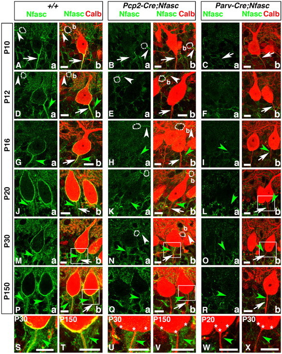
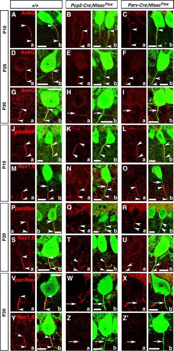
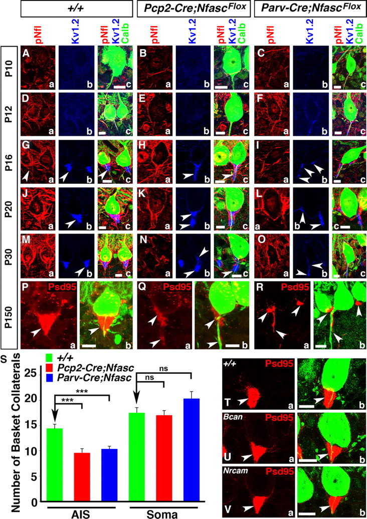
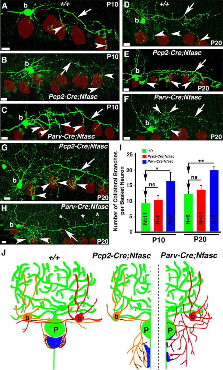
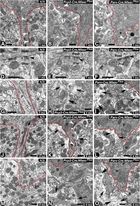
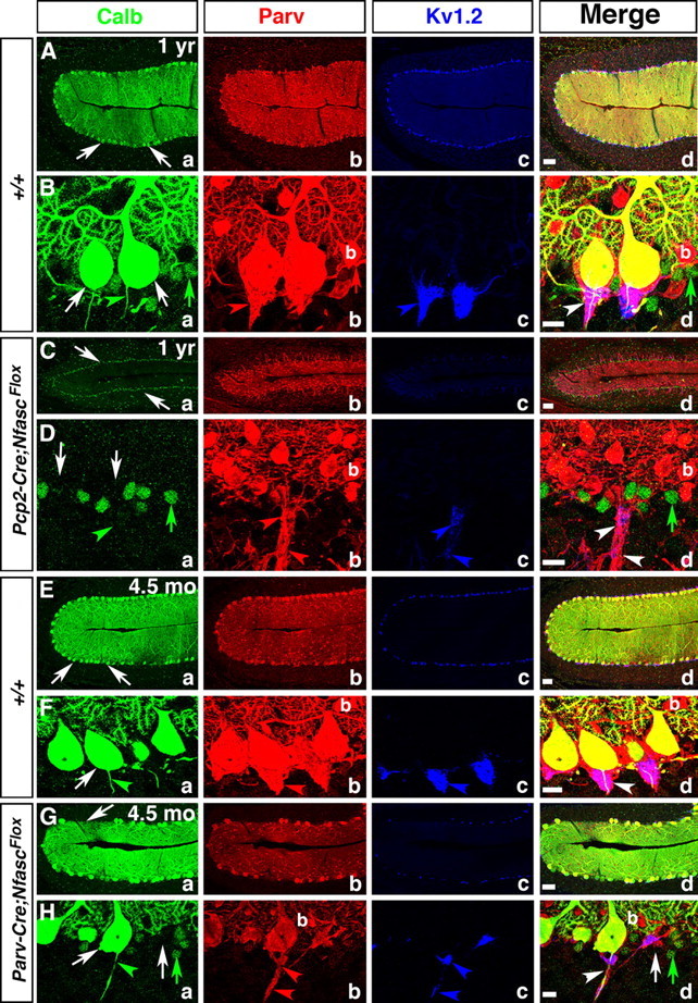
Basket axon collaterals synapse onto the Purkinje soma/axon initial segment (AIS) area to form specialized structures, the pinceau, which are critical for normal cerebellar function. Mechanistic details of how the pinceau become organized during cerebellar development are poorly understood. Loss of cytoskeletal adaptor protein Ankyrin G (AnkG) results in mislocalization of the cell adhesion molecule Neurofascin (Nfasc) at the Purkinje AIS and abnormal organization of the pinceau. Loss of Nfasc in adult Purkinje neurons leads to slow disorganization of the Purkinje AIS and pinceau morphology. Here, we used mouse conditional knock-out techniques to show that selective loss of Nfasc, specifically in Purkinje neurons during early development, prevented maturation of the AIS and resulted in loss of Purkinje neuron spontaneous activity and pinceau disorganization. Loss of Nfasc in both Purkinje and basket neurons caused abnormal basket axon collateral branching and targeting to Purkinje soma/AIS, leading to extensive pinceau disorganization, Purkinje neuron degeneration, and severe ataxia. Our studies reveal that the Purkinje Nfasc is required for AIS maturation and for maintaining stable contacts between basket axon terminals and the Purkinje AIS during pinceau organization, while the basket neuron Nfasc in combination with Purkinje Nfasc is required for proper basket axon collateral outgrowth and targeting to Purkinje soma/AIS. Thus, cerebellar pinceau organization requires coordinated mechanisms involving specific Nfasc functions in both Purkinje and basket neurons.


Figures

Figure 1.
Figure 1.
Molecular organization of the cerebellar pinceau during postnatal development. Wild-type cerebellar sections from ages P10–P90 (indicated on left) and immunostained against Parv, Calb, pNfl, and KV1.2. A, B, At P10, very few basket neuron (b) collaterals have reached Purkinje neuron (P) AISs (arrows). C, D, At P12, a few basket neuron (b) collaterals have arrived at the Purkinje AIS (arrowhead) and faint traces of KV1.2 channels can be seen around the Purkinje AIS. E, F, By P16, more basket neuron (b) collaterals surround the Purkinje (P) AIS, and KV1.2 channels are enriched at the basket neuron terminals. G, H, At P20, the cone-shape of the pinceau is formed, as evident by Parv (Gb, arrow), pNfl (Hb, arrowhead; Hd, arrow), and KV1.2 clustering (Hc, arrowhead). I, J, At P30, the pinceau is fully mature (Ib, Jd, arrows; Jb, Jc arrowheads). K, L, At P90, the structure of the pinceau does not display major anatomical changes compared withP30 (Kb, Ld, arrows; Lb, Lc, arrowheads). M–Q, Cerebellar sections from Gad67-GFP BAC transgenic mice at ages P10 (M), P12 (N), P16 (O), P20 (P), and P30 (Q) immunostained against GFP (green) and Calb (red). The main basket axons (arrows) give off collaterals (arrowheads) that extend toward the Purkinje AIS begining at P10 (M, arrowhead) and form the cone-shape pinceau at P30 (Q, arrowhead). R, Representative drawings of each stage of pinceau development. Purkinje neurons (P) in green, basket neurons (b) in red and orange and potassium channels in blue. Scale bars: 10 μm.
Figure 2.
Figure 2.
Purkinje and basket-specific ablation of Neurofascin reveals its localization to both Purkinje and basket neurons. Wild-type, Pcp2-Cre;NfascFlox and Parv-Cre;NfascFlox cerebellar sections from ages P10–P150 as indicated on left (A–R) or within panel (S–X), and immunostained against Nfasc (a, green) and Calb (b, red). Note that in P10 wild-type cerebella, Nfasc is localized to the Purkinje AIS (A, arrows) and basket (b) AIS (A, white arrowheads). At later stages, in wild-type cerebella, Nfasc is localized around the entire Purkinje soma and AIS (D, G, J, M, P, arrows), as well as the basket AIS (D, white arrowhead). Nfasc is also localized to basket axon terminals as they form the pinceau in wild-type cerebella (D, G, J, M, P, green arrowheads). In Pcp2-Cre;NfascFlox cerebella, Nfasc is ablated from the Purkinje soma/AIS (B, E, H, K, N, Q, arrows) but remains at the basket AIS (B, E, H, N, white arrowhead) and pinceau (H, K, N, Q, green, arrowheads). In Parv-Cre;NfascFlox cerebella, Nfasc is lost from the Purkinje soma/AIS (C, F, I, L, O, R, arrows), basket AIS, and pinceau (I, L, O, R, green arrowheads). Higher magnification shows that Nfasc is present at the Purkinje soma/AIS and pinceau in wild-type (S, T, green arrowheads), is missing from the Purkinje soma/AIS in Pcp2-Cre;NfascFlox (U, V, stars), while remaining at the pinceau in Pcp2-Cre;NfascFlox (U, V, green arrowheads), and is missing from both the Purkinje soma/AIS (stars) and the pinceau (green arrowheads) in Parv-Cre;NfascFlox cerebella (W, X). Scale bars: 10 μm.
Figure 3.
Figure 3.
Early postnatal loss of Nfasc disrupts Purkinje AIS maturation and stabilization. Wild-type, Pcp2-Cre;NfascFlox and Parv-Cre;NfascFlox cerebellar sections from mice ages P10–P30 immunostained against AnkG (A–Ia, red), panNaV (J–L; P–R, V–Xa, red), NaV1.6 (M–O; S–U, Y–Z'a, red), and Calb (A–Z'b, green). AnkG is localized (between arrowheads) to the wild-type Purkinje AIS at P10, P20, and P30. In both mutants, AnkG remains (between arrowheads) at the Purkinje AIS at P10, moves distally down the axon at P20 (E, F, between arrowheads), and is undetectable from the Purkinje axon by P30 (H, I, arrows). At P10 (J, between arrowheads), P20 (P, between arrowheads), and P30 (V, between arrowheads), panNaV shows AIS localization in wild-type Purkinje AIS. Localization of panNaV remains the same as in the wild-type in both mutants at P10 (K, L, between arrowheads), but at P20 panNaV shows severely reduced immunostaining at the mutant AISs (Q, R, between arrowheads), and is completely missing from the P30 mutant AISs (W, X, arrows). Localization of NaV1.6 at the wild-type AIS at P10 shows that NaV1.6 is located distally in the AIS (M, between arrowheads) and moves to fill the entire AIS at P20 and P30 (S, Y, between arrowheads). Localization of NaV1.6 remains the same as wild-type in both mutants at P10 (N, O, between arrowheads) and remains distally localized at P20 (T, U, between arrowheads), and is undetectable at the AIS at P30 (Z, Z', arrows). Scale bars: 10 μm.
Figure 4.
Figure 4.
Ablation of Nfasc disrupts basket axon targeting and pinceau organization. Wild-type (A, D, G, J, M), Pcp2-Cre;NfascFlox (B, E, H, K, N), and Parv-Cre;NfascFlox (C, F, I, L, O) cerebellar sections from ages P10 (A–C), P12 (D–F), P16 (G–I), P20 (J–L), and P30 (M–O) immunostained against pNfl (a, red), KV1.2 (b, blue), and Calb (c, green, merged). Pinceau disruption becomes evident in Pcp2-Cre;NfascFlox cerebella at P16, when fewer collaterals appear to reach the Purkinje AIS (Ha, c), and KV1.2 does not become as enriched as at the wild-type pinceau (Hb, c). This trend continues at P20 and P30, where fewer collaterals reach the Purkinje AIS (Ka, Na) and potassium channels fail to cluster properly and appear as broken clusters compared with wild-type (Kb, Nb). In Parv-Cre;NfascFlox cerebella at P16, basket axon targeting is disrupted (Ia, c) and KV1.2 clusters ectopically around the soma and at random places along the Purkinje AIS (Ib, c). This disruption continues at P20 and P30 in Parv-Cre;NfascFlox cerebella with altered basket axon targeting (La, Oa) and ectopic KV1.2 clusters (Lb, Ob). Wild-type (P, T), Pcp2-Cre;NfascFlox (Q), Parv-Cre;NfascFlox (R), Brevican knock-out (Bcan, U), and NrCAM knock-out (V) cerebella immunostained against PSD95 (a, red) and Calb (b, green, merged). At P150, the wild-type pinceau maintains its cone-shaped PSD95 localization (P, T), while it is completely disrupted in both Pcp2-Cre;NfascFlox (Q) and Parv-Cre;NfascFlox cerebella (R). S, Quantification of basket axon collateral targeting to the Purkinje AIS and soma in Pcp2-Cre;NfascFlox and Parv-Cre;NfascFlox cerebella. The pinceau remains intact in both Bcan (U) and NrCAM (V) mutants. Scale bars: 10 μm. ns, Not significant. ***p < 0.001.
Figure 5.
Figure 5.
Loss of Nfasc in basket neurons causes mistargeting of basket axon collaterals. Wild-type (A, D), Pcp2-Cre;NfascFlox, (B, E, G), and Parv-Cre;NfascFlox (C, F, H) cerebellar sections from ages P10 (A–C) and P20 (D–H) in which basket cells were injected with biocytin and immunostained with Alexa-conjugated Streptavidin (green). Note that basket axons project toward the Purkinje neuron layer in Pcp2-Cre;NfascFlox mutants, but branch excessively at their terminals (B, E, G, arrowheads). In Parv-Cre;NfascFlox mutants, basket axons branch excessively from the beginning of the extension (C, F, H, arrowheads) and sometimes point in opposite directions (H, asterisks). Purkinje somas are outlined as red circles. I, Quantification of the number of primary basket terminal branches coming off the main basket axon branch at each time point reveal an increase in branching along the length of the Parv-Cre;NfascFlox basket axon compared with wild-type and Pcp2-Cre;NfascFlox basket neurons. J, Representative drawings summarizing the pinceau structures observed in wild-type, Pcp2-Cre;NfascFlox and Parv-Cre;NfascFlox cerebella. Scale bars: 10 μm. ns, Not significant. *p < 0.05; **p < 0.01.
Figure 6.
Figure 6.
Cerebellar pinceau disorganization and infiltration of glial processes in Nfasc-deficient Purkinje and basket neuron cerebella. A–C, Electron micrographs of the pinceau region of cerebellar sections from one-month-old wild-type (+/+) pinceau show compact, electron-dense basket axon collaterals (B) surrounding the Purkinje soma/AIS highlighted by red line (A). Note the presence of synapses between Purkinje (P) AIS and basket collaterals (B) (A, arrowheads). In 1-month-old Pcp2-Cre;NfascFlox cerebella, the pinceau appears slightly disorganized, with spaces beginning to form between basket axon terminals (B) (B, arrows). Note the presence of a synapse between the basket terminal and the Purkinje soma (B, arrowhead). In 1-month-old Parv-Cre;NfascFlox cerebella, the pinceau appears more disorganized with glial processes infiltrating spaces between basket axon terminals (C, arrows). D–F, Electron micrographs at higher magnification showing synapses (black arrowheads) with presynaptic (basket terminal) and postsynaptic (Purkinje AIS) areas in the wild-type (D), Pcp2-Cre;NfascFlox (E), and Parv-Cre;NfascFlox (F) cerebella. G–I, Electron micrographs from cerebellar sections from 3-month-old wild-type cerebella show highly compact basket terminals surrounding the Purkinje AIS (G). Pcp2-Cre;NfascFlox pinceau at 3 months shows increased infiltration of glial processes (H, black arrows). A 3-month-old Parv-Cre;NfascFlox pinceau shows increased disorganization of basket terminals surrounding the Purkinje soma/AIS (I, black arrows). J–O, Electron micrographs of the pinceau from 4-month-old wild-type (J, M), Pcp2-Cre;NfascFlox (K, N), and Parv-Cre;NfascFlox (L, O) cerebella. Wild-type pinceau show compact basket axon terminals surrounding the Purkinje soma/AIS. The Pcp2-Cre;NfascFlox and Parv-Cre;NfascFlox cerebella show severely disorganized basket terminals with increased infiltrating glial processes (black arrows). In some instances, in Parv-Cre;NfascFlox cerebella the entire pinceau areas are completely disrupted (O, black arrows). Scale bars: 0.5 μm.
Figure 7.
Figure 7.
Purkinje neuron-specific ablation of Nfasc abolishes Purkinje neuron spontaneous activity and reduces inhibitory inputs to Purkinje neurons. A, Representative traces of spontaneous Purkinje neuron firing in wild-type (+/+) and Pcp2-Cre;NfascFlox Purkinje neurons. B, Plot of the spontaneous firing rates of wild-type (black circles) and Pcp2-Cre;NfascFlox mutant (white circles) Purkinje neurons. Larger diamonds show average spontaneous firing rates of Purkinje neurons in wild-type (black diamond) and Pcp2-Cre;NfascFlox mutants (white diamond). C, Representative traces of the action potential train response of Purkinje neurons to injected current at increasing depolarizing steps. D–F, Output responses from wild-type (black circles) and Pcp2-Cre;NfascFlox (white circles) Purkinje neurons to increasing levels of injected current. G, Representative traces of mIPSC recordings from wild-type and Pcp2-Cre;NfascFlox Purkinje neurons. H, Plot of mIPSC frequency reveals decreased frequency in Pcp2-Cre;NfascFlox mutant Purkinje neurons. I, Plot of mIPSC amplitude revealing no change between the wild-type and Pcp2-Cre;NfascFlox mutant Purkinje neurons.
Figure 8.
Figure 8.
Ablation of Nfasc in Purkinje neurons leads to progressive Purkinje neuron degeneration. One-year old wild-type (A, B), one-year-old Pcp2-Cre;NfascFlox (C, D), 4.5-month-old wild-type (E, F), and 4.5-month-old Parv-Cre;NfascFlox (G, H) cerebellar sections immunostained against Calb (a, green), Parv (b, red), KV1.2 (c, blue), and merged (d). One-year and 4.5 month wild-type cerebella maintain a full complement of healthy Purkinje neuron layer (Aa, Ba, Ea, Fa, arrows), while one-year-old Pcp2-Cre;NfascFlox cerebella show decreased molecular layer size with loss of Purkinje neurons (Ca, Da, arrows). The pinceau is stably maintained in one-year and 4.5-month-old wild-type cerebella (Bb–d, Fb–d, arrows). In Pcp2-Cre;NfascFlox cerebella, the basket axons appear clumped together (Db, d, arrowheads), but do not form a pinceau structure as all Purkinje neurons have died (Da, arrows). In 4.5 month Parv-Cre;NfascFlox cerebella, some Purkinje cells have begun to degenerate (Ga, d; Ha, d, arrows) and the pinceau structure remains disrupted with ectopic clusters of potassium channels (Hb–d, arrowheads). Scale bars: (in d) A, C, E, G, 50 μm; B, D, F, H, 10 μm.

Similar articles

Cited by

References

    1. Ango F, di Cristo G, Higashiyama H, Bennett V, Wu P, Huang ZJ. Ankyrin-based subcellular gradient of neurofascin, an immunoglobulin family protein, directs GABAergic innervation at purkinje axon initial segment. Cell. 2004;119:257–272. - PubMed
    1. Barski JJ, Dethleffsen K, Meyer M. Cre recombinase expression in cerebellar Purkinje cells. Genesis. 2000;28:93–98. - PubMed
    1. Bastianelli E. Distribution of calcium-binding proteins in the cerebellum. Cerebellum. 2003;2:242–262. - PubMed
    1. Bennett V, Baines AJ. Spectrin and ankyrin-based pathways: metazoan inventions for integrating cells into tissues. Physiol Rev. 2001;81:1353–1392. - PubMed
    1. Benton MD, Raman IM. Stabilization of Ca current in Purkinje neurons during high-frequency firing by a balance of Ca-dependent facilitation and inactivation. Channels. 2009;3:393–401. - PMC - PubMed

Publication types

MeSH terms

LinkOut - more resources