Review
. 2012 May;139(9):1535-46.
doi: 10.1242/dev.069963.

Transcriptional co-regulation of neuronal migration and laminar identity in the neocortex

Affiliations
Review

Transcriptional co-regulation of neuronal migration and laminar identity in the neocortex

Kenneth Y Kwan et al. Development. 2012 May.

Abstract

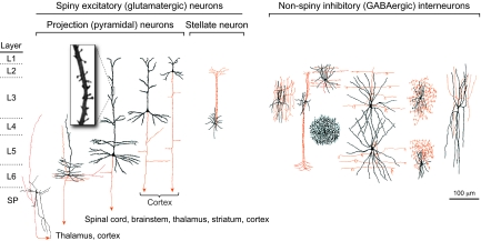
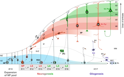
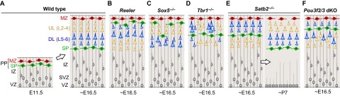
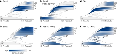
The cerebral neocortex is segregated into six horizontal layers, each containing unique populations of molecularly and functionally distinct excitatory projection (pyramidal) neurons and inhibitory interneurons. Development of the neocortex requires the orchestrated execution of a series of crucial processes, including the migration of young neurons into appropriate positions within the nascent neocortex, and the acquisition of layer-specific neuronal identities and axonal projections. Here, we discuss emerging evidence supporting the notion that the migration and final laminar positioning of cortical neurons are also co-regulated by cell type- and layer-specific transcription factors that play concomitant roles in determining the molecular identity and axonal connectivity of these neurons. These transcriptional programs thus provide direct links between the mechanisms controlling the laminar position and identity of cortical neurons.


Figures

Fig. 1.
Fig. 1.
Major neuronal cell types of the adult cerebral cortex. Cortical neurons (shown here for primates) are categorized into two major classes: spiny excitatory (glutamatergic) neurons (left panel) and non-spiny inhibitory (GABAergic) interneurons (right panel). The former, the dendrites of which are decorated by numerous post-synaptic membranous protrusions termed spines (see inset), include the projection (pyramidal) neurons, the principal cells of the neocortex, and stellate neurons (see Glossary, Box 1), which are mostly found in L4 of primary sensory areas. Projection neurons display marked layer- and subtype-specific differences in the morphology of their dendrites (black) and in the targets of their axonal projections (red); neurons within the deep layers (L5 and L6) and the subplate (SP) project axons that target cortex and subcortical structures, including striatum, thalamus, brainstem and spinal cord, whereas upper-layer (L2-L4) neurons project axons within the cortex. The non-spiny interneurons, which are highly diverse in morphology, neurochemistry and electrophysiology, project axons within a local circuit. Subtypes of interneurons also display laminar preferences, thereby contributing to layer differences in cortical circuitry. Adapted from Jones (Jones, 1986).
Fig. 2.
Fig. 2.
Schematic of projection neuron generation and migration in the mouse neocortex. Prior to the onset of neurogenesis, neural progenitors (NPs) in the ventricular zone (VZ; blue) of the developing neocortex divide symmetrically to expand the progenitor pool, undergoing interkinetic nuclear migration (IKNM) as they progress through the cell cycle. Starting at ∼E11.5, NPs assume radial glial morphology and begin dividing asymmetrically to generate neurons, which migrate from the germinal zones guided by radial glia cells (RGC) to reach the mantle layers. The first projection neurons settle within the preplate (PP) to form the nascent cortical plate (CP), which will subsequently become layers (L) 2 to 6 of the neocortex. Additional incoming CP neurons then split the PP into the marginal zone (MZ) and the subplate (SP). As neurogenesis progresses, diverse subtypes of projection neurons are generated sequentially through successive asymmetric divisions of NPs. Thus, neurons destined for the SP are generated first, followed by those destined for the deep layers (L6 and L5; red), and finally, those destined for the upper layers (L4, L3 and L2; green). The migration of newborn neurons into the CP occurs in an inside-first, outside-last manner; early-born neurons form the deep layers, whereas later-born neurons migrate past older neurons to form more superficial layers. Therefore, the cortical layers are sequentially generated in an ‘inside-out’ fashion. Some daughter cells of NPs become intermediate progenitor cells (IPCs), migrating away from the VZ and undergoing symmetric neurogenic divisions in the SVZ. This mode of neurogenesis contributes significantly to upper layer neurons. At the end of neurogenesis at ∼E17.5, the radial scaffold is dismantled and NPs become gliogeneic, generating cortical and subependymal zone (SEZ) astrocytes (Ast) and giving rise to a layer of ependymal cells (EL). The tangential migration and laminar positioning of interneurons are not illustrated. BV, blood vessel; CR, Cajal-Retzius neuron; DL Pyr, deep-layer pyramidal neuron; IZ, intermediate zone; UL Pyr, upper-layer pyramidal neuron, WM, white matter.
Fig. 3.
Fig. 3.
Summary of neocortical neuronal migration and positioning defects in knockout mice. (A) Migration and laminar positioning in wild-type mouse. (B) In the cortex of Reeler mutants, preplate (PP) splitting is defective and lamination of the subplate (SP) and the entire cortical plate (CP) (L6-L2) is inverted. (C) In the Sox5-deficient cortex, the PP also fails to segregate but only the deep layers (DL) are inverted. The upper layers (UL) are formed without overt defect. (D) In the Tbr1-deficient cortex, the PP is partially split but the SP is mis-positioned to the middle of the CP. Deep-layer neurons form abnormal clusters and the majority of upper-layer neurons fail to migrate past the ectopic SP. (E) In the Satb2-deficient cortex, the migration of late-born neurons is delayed. This defect, however, is corrected within the first postnatal week. (F) In the Pou3f2/3 double knockout (dKO) neocortex, the PP is split and SP and L6 are formed without abnormality. The migration of L5-L2 projection neurons, however, is stalled below the SP. IZ, intermediate zone; MZ, marginal zone; P, postnatal day; SVZ, subventricular zone; VZ, ventricular zone.
Fig. 4.
Fig. 4.
Summary of spatiotemporal expression of transcription factors co-regulating neuronal migration and identity in the mouse neocortex. (A) Sox5 is specifically expressed in post-mitotic neurons and is absent from ventricular zone (VZ) and subventricular zone (SVZ) progenitors. From embryonic development until the perinatal period, its expression is specific to the deep layers, being strongly present in L6 and subplate (SP) neurons and in some L5 neurons. After the first postnatal week, some upper layer neurons also express Sox5. (B) Fezf2 is highly expressed in early VZ progenitors and their subcortically projecting deep-layer neuron progenies, but is absent from the upper-layer neurons and the late progenitors from which they are derived. In late embryonic development, Fezf2 is post-mitotically downregulated in L6 neurons, thereby giving rise to its L5-enriched postnatal pattern. (C) Tbr1 is specifically expressed in post-mitotic corticothalamic neurons of L6 and the SP, from embryonic development through to the first postnatal week. Thereafter, some upper layer neurons also turn on Tbr1 expression. (D) Satb2 is highly expressed in post-mitotic corticocortical L2-L5 neurons, beginning as these neurons migrate through the intermediate zone (IZ) and persisting postnatally. It is absent from subcortical projection neurons. (E,F) Pou3f2 and Pou3f3 are expressed in late progenitors and in their L2-L5 neuron progenies. Pou3f2 and Pou3f3 expression persists in these neurons, from their generation and migration, through their post-migratory differentiation and beyond.

References

    1. Akbarian S., Kim J. J., Potkin S. G., Hetrick W. P., Bunney W. E., Jones E. G. (1996). Maldistribution of interstitial neurons in prefrontal white matter of the brains of schizophrenic patients. Arch. Gen. Psychiatry 53, 425–436 - PubMed
    1. Alcamo E. A., Chirivella L, Dautzenberg M, Dobreva G, Fariñas I, Grosschedl R., McConnell S. K. (2008). Satb2 regulates callosal projection neuron identity in the developing cerebral cortex. Neuron 57, 364–377 - PubMed
    1. Alfano C., Viola L., Heng J. I., Pirozzi M., Clarkson M., Flore G., De Maio A., Schedl A., Guillemot F., Studer M. (2011). COUP-TFI promotes radial migration and proper morphology of callosal projection neurons by repressing Rnd2 expression. Development 138, 4685–4697 - PubMed
    1. Allendoerfer K. L., Shatz C. J. (1994). The subplate, a transient neocortical structure: its role in the development of connections between thalamus and cortex. Annu. Rev. Neurosci. 17, 185–218 - PubMed
    1. Anderson S. A., Volk D. W., Lewis D. A. (1996). Increased density of microtubule associated protein 2-immunoreactive neurons in the prefrontal white matter of schizophrenic subjects. Schizophr. Res. 19, 111–119 - PubMed

Publication types

Substances