. 2012 May;139(9):1630-9.
doi: 10.1242/dev.075564.

COUP-TFII controls amygdala patterning by regulating neuropilin expression

Affiliations

COUP-TFII controls amygdala patterning by regulating neuropilin expression

Ke Tang et al. Development. 2012 May.

Abstract

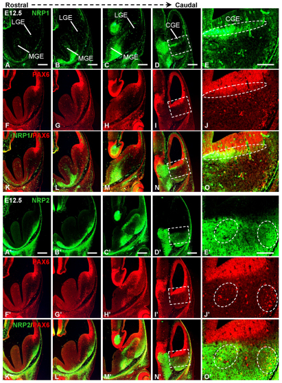
The development of the progenitor zones in the pallium, lateral ganglionic eminence (LGE) and medial ganglionic eminence (MGE) in the subpallium has been well studied; however, so far the role of the caudal ganglionic eminence (CGE), a posterior subpallial domain, in telencephalon patterning remains poorly understood. COUP-TFII, an orphan nuclear receptor, is preferentially expressed in the CGE. We generated COUP-TFII mouse mutants, using Rx-Cre (RxCre;COUP-TFII(F/F)), to study its function in telencephalon development. In these mutants, we found severe defects in the formation of the amygdala complex, including the lateral (LA), basolateral (BLA) and basomedial (BMA) amygdala nuclei. Molecular analysis provided evidence that the migration of CGE-derived Pax6(+) cells failed to settle into the BMA nucleus, owing to reduced expression of neuropilin 1 (Nrp1) and Nrp2, two semaphorin receptors that regulate neuronal cell migration and axon guidance. Our ChIP assays revealed that Nrp1 and Nrp2 genes are the direct targets of COUP-TFII in the telencephalon in vivo. Furthermore, our results showed that the coordinated development between the CGE originated subpallial population (Pax6(+) cells) and pallial populations (Tbr1(+) and Lhx2(+) cells) was essential for patterning the amygdala assembly. Our study presented novel genetic evidence that the caudal ganglionic eminence, a distinct subpallial progenitor zone, contributes cells to the basal telencephalon, such as the BMA nucleus.


Figures

Fig. 1.
Fig. 1.
COUP-TFII expression in the CGE and the developing mature amygdala neurons. (A-H) COUP-TFII expression at E12.5. Series of the coronal sections along the rostrocaudal axis are shown in A-D. (E-H) Higher magnification of the boxes in A-D. (I-L) Pax6 expression at E12.5. Open arrowheads in I-K indicate that the Pax6+ cells originated from the dLGE. Arrowheads in L indicate two Pax6+ migration streams derived from the CGE. (M-P) Double immunostaining of COUP-TFII and Pax6. Insets show the higher magnification images at regions 1 and 2 in P. (Q-T) Tbr1 expression at E12.5. (U-X) Double immunostaining of COUP-TFII and Tbr1. sLP/VP at the caudal telencephalon probably corresponds to the primordia of the pallial amygdala, including the LA and BLA nuclei. (Y1-5) COUP-TFII expression in the amygdala complex at P0. (Z1-5) COUP-TFII expression in the amygdala complex at 1 month. Amy, amygdala anlage; CGE, caudal ganglionic eminence; CH, Cortical hen; CP, choroids plexus; CVCo, mantle zone of caudoventral cortex; dLGE, dorsal lateral ganglionic eminence; LGE, lateral ganglionic eminence; MGE, medial ganglionic eminence; sDP, dorsal pallium originated Tbr1+ stream; sLP/VP, lateral pallium/ventral pallium originated Tbr1+ stream. Scale bars: 200 μm in A-D; 100 μm in E-X, Y1-5 and Z1-5; 500 μm in the low-magnification image in Y; 1 mm in the montage image at 1 month (1M) in Z.
Fig. 2.
Fig. 2.
The formation of the amygdala is compromised morphologically in the COUP-TFII mutant at 3 months of age postnatally. Nissl staining of brains sections from the control and mutant mice at 3 months of age. (A-F) Low-magnification images at amygdala complex from the control (A,C,E) and mutant mouse (B,D,F). (G-L) High-magnification images at the BMA area from the control (G,I,K) and mutant mouse (H,J,L). BLA, basolateral amygdala nucleus; BMA, basomedial amygdala nucleus; LA, lateral amygdala nucleus. Scale bars: 500 μm in A-F; 200 μm in G-L.
Fig. 3.
Fig. 3.
The abnormal basolateral complex in the COUP-TFII mutant mouse at P1. Coronal sections at four planes along the rostrocaudal axis of the amygdala from the heterozygous control mouse (RxCre;F/+) and mutant mouse (RxCre;F/F) were used to perform immunohistochemical assays. (A-D′) COUP-TFII expression in the control (A-D) and mutant (A′-D′) at P1. (E-H′) Mef2C expression in the control (E-H) and mutant (E′-H′) at P1. (I-L′) Tbr1 expression in the control (I-L) and mutant (I′-L′) at P1. (M-P′) lacZ expression in the control (M-P) and mutant (M′-P′) at P1. BLA, basolateral amygdala nucleus; BMA, basomedial amygdala nucleus; CeA, center amygdala nucleus; LA, lateral amygdala nucleus. Scale bars: 200 μm.
Fig. 4.
Fig. 4.
The expression profiles of COUP-TFII, Pax6 and Tbr1 in the ventral telencephalon at E12.5. (A-H) COUP-TFII expression in the control (A-D) and mutant (E-H) at E12.5. (I-P) Pax6 expression in the control (I-L) and mutant (M-P) at E12.5. (L′,P′) Insets in L,P. (Q-X) Tbr1 expression in the control (Q-T) and mutant (U-X) at E12.5. Arrows and arrowheads indicate sLP/VP and sDP migrating streams in the CGE, respectively. CGE, caudal ganglionic eminence; dLGE, dorsal lateral ganglionic eminence; LGE, lateral ganglionic eminence; MGE, medial ganglionic eminence; sDP, dorsal pallium originated Tbr1+ stream; sLP/VP, lateral pallium/ventral pallium originated Tbr1+ stream. Scale bars: 200 μm in A-X; 100 μm in L′,P′.
Fig. 5.
Fig. 5.
The expression profiles of Pax6 and Tbr1 at the ventral telencephalon at E13.5. (A-H) Pax6 expression in the control (A,C,E,G) and mutant (B,D,F,H) at E13.5. Arrowheads indicate Pax6+ migratory streams. Arrow indicates abnormally localized Pax6+ cells in the mutant. (I-P) Tbr1 expression in the control (I,K,M,O) and mutant (J,L,N,P) at E13.5. Asterisks indicate Pax6+ cell migration route. Arrowheads indicate multiple Tbr1+ cell layers in the mutant ventral CGE. Arrow indicates ectopic Tbr1+ cells in the mutant BMA locus. BMA, basomedial amgydala nucleus; CGE, caudal ganglionic eminence; dLGE, dorsal lateral ganglionic eminence; sDP, dorsal pallium originated Tbr1+ stream; sLP/VP, lateral pallium/ventral pallium originated Tbr1+ stream. Scale bars: 200 μm.
Fig. 6.
Fig. 6.
Nrp1 and Nrp2 are direct targets of COUP-TFII in embryonic forebrain in vivo. (A) Quantitative real-time PCR assays were conducted to profile the expression of COUP-TFII, Shh, Bmp4, Fgf8, Gsh2, Lhx2, Lhx5, Lhx9, Dbx1, Otx2, Epharin5a, Sfrp2, Sema3a, Sema3f, Nrp1 and Nrp2 in the control (n=3) and mutant (n=4). Student’s t-test, *P<0.05, ***P<0.005. (Ba-d) Immunohistochemical assays with Nrp1 antibody at E12.5 and E13.5. (Ca-d) Immunohistochemical assays with Nrp2 antibody at E12.5 and E13.5. CGE, caudal ganglionic eminence. (D,E) In vivo ChIP assays with chromatin prepared from the forebrains at E12.5. Student’s t-test, **P<0.01, ***P<0.005, P<0.001. Data are mean±s.e.m. Scale bars: 300 μm in Ba,Bb,Ca,Cb; 200 μm in Bc,Bd,Cc,Cd.
Fig. 7.
Fig. 7.
Nrp1 and Nrp2 are localized in the Pax6+ pre-migratory cells or migratory cells in the CGE, but not in the Pax6+ migratory cells derived from the dLGE. (A-D) Nrp1 expression in the ventral telencephalon at E12.5. (E) Inset in D. (F-I,F-I′) Pax6 expression at E12.5. (J) Inset in I; (J′) inset in I′. (K-N) Pax6 and Nrp1 co-immunostaining. (O) Inset in N. (A′-D′) Nrp2 expression in the ventral telencephalon at E12.5. (E′) Inset in D′. (K′-N′) Pax6 and Nrp2 co-immunostaining. (O′) Inset in N′. CGE, caudal ganglionic eminence; LGE, lateral ganglionic eminence; MGE, medial ganglionic eminence. Scale bars: 200 μm in A-D,A’-D’,F-I,F’-I’,K-N,K’-N’; 100 μm in E,E′,J,J′,O,O’.

Similar articles

Cited by

References

    1. Bulchand S., Grove E. A., Porter F. D., Tole S. (2001). LIM-homeodomain gene Lhx2 regulates the formation of the cortical hem. Mech. Dev. 100, 165–175 - PubMed
    1. Bulfone A., Smiga S. M., Shimamura K., Peterson A., Puelles L., Rubenstein J. L. (1995). T-brain-1: a homolog of Brachyury whose expression defines molecularly distinct domains within the cerebral cortex. Neuron 15, 63–78 - PubMed
    1. Bupesh M., Legaz I., Antonio Abellán A., Medina L. (2011a). Multiple telencephalic and extratelencephalic embryonic domains contribute neurons to the medial extended amygdala. J. Comp. Neurol. 519, 1505–1525 - PubMed
    1. Bupesh M., Abellán A., Medina L. (2011b). Genetic and experimental evidence supports the continuum of the central extended amygdala and a mutiple embryonic origin of its principal neurons. J. Comp. Neurol. 519, 3507–3531 - PubMed
    1. Butt S. J., Fuccillo M., Nery S., Noctor S., Kriegstein A., Corbin J. G., Fishell G. (2005). The temporal and spatial origins of cortical interneurons predict their physiological subtype. Neuron 48, 591–604 - PubMed

Publication types

MeSH terms