. 2012 May;139(9):1651-61.
doi: 10.1242/dev.068395.

Dynamic in vivo binding of transcription factors to cis-regulatory modules of cer and gsc in the stepwise formation of the Spemann-Mangold organizer

Affiliations

Dynamic in vivo binding of transcription factors to cis-regulatory modules of cer and gsc in the stepwise formation of the Spemann-Mangold organizer

Norihiro Sudou et al. Development. 2012 May.

Abstract

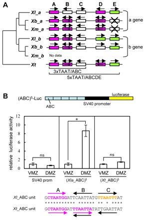
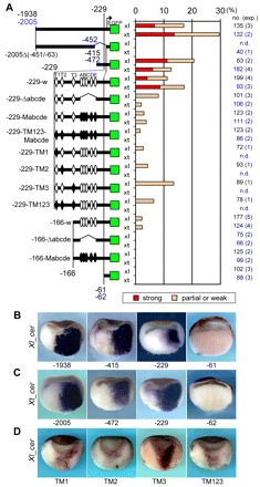
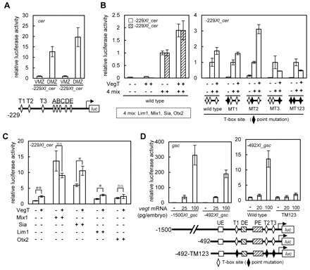
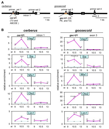
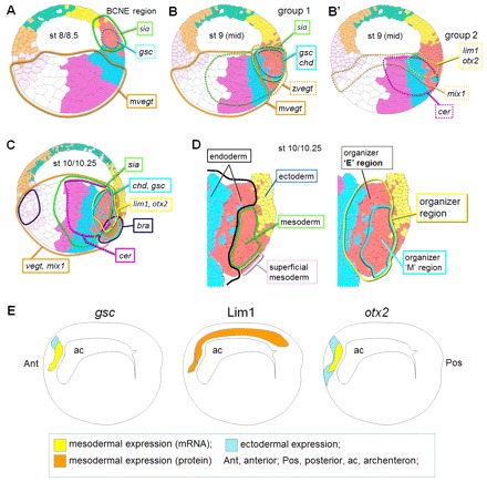
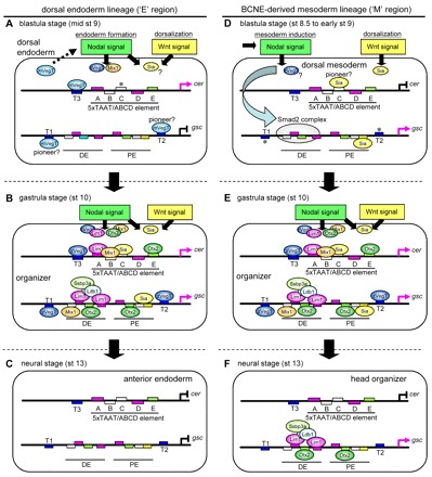
How multiple developmental cues are integrated on cis-regulatory modules (CRMs) for cell fate decisions remains uncertain. The Spemann-Mangold organizer in Xenopus embryos expresses the transcription factors Lim1/Lhx1, Otx2, Mix1, Siamois (Sia) and VegT. Reporter analyses using sperm nuclear transplantation and DNA injection showed that cerberus (cer) and goosecoid (gsc) are activated by the aforementioned transcription factors through CRMs conserved between X. laevis and X. tropicalis. ChIP-qPCR analysis for the five transcription factors revealed that cer and gsc CRMs are initially bound by both Sia and VegT at the late blastula stage, and subsequently bound by all five factors at the gastrula stage. At the neurula stage, only binding of Lim1 and Otx2 to the gsc CRM, among others, persists, which corresponds to their co-expression in the prechordal plate. Based on these data, together with detailed expression pattern analysis, we propose a new model of stepwise formation of the organizer, in which (1) maternal VegT and Wnt-induced Sia first bind to CRMs at the blastula stage; then (2) Nodal-inducible Lim1, Otx2, Mix1 and zygotic VegT are bound to CRMs in the dorsal endodermal and mesodermal regions where all these genes are co-expressed; and (3) these two regions are combined at the gastrula stage to form the organizer. Thus, the in vivo dynamics of multiple transcription factors highlight their roles in the initiation and maintenance of gene expression, and also reveal the stepwise integration of maternal, Nodal and Wnt signaling on CRMs of organizer genes to generate the organizer.


Figures

Fig. 1.
Fig. 1.
Comparison of cer promoter sequences and homeodomain binding sites between Xenopus species. (A) Evolutionary conservation of ABCDE elements between Xenopus species. The ABCDE element is schematically aligned for two homoeologous genes a and b of the X. laevis (Xl), X. borealis (Xb) and X. muelleri (Xm) cer genes together with the single cer gene of X. tropicalis (Xt). Phylogenetic relationships were determined by ClustalW using 5′ and exon sequences (supplementary material Fig. S1C) (DDBJ/Genbank Accession Numbers: Xl_cer-a, AB086394; Xl_cer-b, AB698643; Xb_cer-a, AB698644; Xb_cer-b, AB698645; Xm_cer-a, AB698642). Magenta boxes indicate Lim1-binding sites (YTAATNN; Y=T or C, NN=TA, TG, CA, or GG), green boxes indicate bicoid sites (TAATCY); white boxes indicate other TAATNN sites. Arrows above and in the box indicate the direction of TAAT (the double-headed arrow indicates the TAATTA sequence; boxes with a cross indicate disrupted TAAT-binding site). (B) Comparison of enhancer activity of the ABC elements between X. laevis and X. tropicalis cer genes. (Xla_ABC)5/Luc and (Xt_ABC)5/Luc are five tandemly repeated ABC elements fused to the SV40 minimal promoter (SV40 prom). Reporter DNA (50 pg/embryo) was injected into the prospective dorsal or ventral marginal zone (DMZ or VMZ, respectively) and luciferase activity was assayed at the gastrula stage (stage 10.5). Data are mean±s.d.; *P<0.05; ns, not significant.
Fig. 2.
Fig. 2.
Cis-element analysis of X. laevis and X. tropicalis cer promoter regions using transgenic reporter genes. (A) Deletion and point mutations of cer promoter-EGFP reporter constructs and their dorsal expression at the gastrula stage. Constructs of Xl_cer and Xt_cer promoter regions are schematically shown on the left, and numbers indicate positions from the transcription start site, whereas numbers in blue indicate position of Xt_cer when different from those of Xl_cer. A bent arrow indicates the transcription start site. Fixed embryos were bisected along the dorsal midline and whole-mount in situ hybridization was performed for EGFP mRNA. Dorsal expression of reporter genes was categorized into strong (orange) and partial or weak (pale orange) expression patterns (supplementary material Fig. S3A), and percentage incidences of each category are presented by bar graphs. The total number of embryos examined and those of experiments in the parentheses are indicated on the right. n.d., not determined. (B,C) Whole-mount in situ hybridization analysis of reporter expression. Typical expression patterns of Xl_cer (B) or Xt_cer (C) reporter constructs are shown. (D) Reporter gene expression of T-box mutant constructs.
Fig. 3.
Fig. 3.
Cooperation of VegT with Mix1, Siamois, Lim1 and Otx2 to activate the cer and gsc enhancers. (A) Dorsal-specific expression of –229Xl_cer and –229Xt_cer as assayed by DNA injection. Experiments were carried out as described in Fig. 1B. (B) Synergy of VegT with a mixture of Mix1, Sia, Lim1 and Otx2. Wild-type constructs of –229Xl_cer (white bar) and –229Xt_cer (diagonally striped bar) (left panel) or T-box mutant constructs of –229Xl_cer (right panel) were co-injected with mRNAs (25 pg mRNA each) for VegT and 4 mix (mix1, sia, lim1 and otx2), as indicated by a plus mark. Mutated T-box sites in –229Xl_cer-MT1, MT2, MT3 and MT123 are indicated by black diamonds, in which TNNCAC was mutated to CNNTAC. (C) Synergy of VegT with Sia and Lim1. The –229Xl_cer construct was co-injected with mRNA as indicated with or without mRNA for VegT. Amounts of injected mRNAs (pg/embryo): vegt, 25; lim1, 25; mix1, 25; sia, 12; otx2, 25. (D) Activation of gsc reporter constructs by VegT. –1500Xl_gsc or –492Xl_gsc was co-injected with vegt mRNA as indicated. The sequence of –1500Xl_gsc has been deposited in DDBJ/Genbank (AB698641).
Fig. 4.
Fig. 4.
ChIP-qPCR analysis for cer-U1 and gsc-U1. (A) Schematic diagrams of gene structures of cer and gsc. Positions of cer-U1 and gsc-U1; primer sets for ChIP-qPCR are indicated. (B) Dynamics of in vivo binding of transcription factors to cer-U1 and gsc-U1. Occupancy of Lim1, Mix1, Sia, Otx2 and VegT proteins at cer-U1 (left panel) and gsc-U1 (right panel) was examined by ChIP-qPCR with primer sets as indicated. qPCR was performed in triplicate (cerberus) or duplicate (goosecoid) unless otherwise indicated in parentheses. Magenta or light-blue lines, specific or preimmune antibodies, respectively, for ChIP-qPCR; st, stage; data are mean±s.e.m.; *P<0.05; **P<0.01. Enrichment of cer-U1 and gsc-U1 regions by ChIP was also examined by PCR/gel electrophoresis analysis.
Fig. 5.
Fig. 5.
Assignment of expression domains of organizer-expressing genes. (A-C) Blastula to gastrula stages. Expression domains at early blastula (A), mid-blastula (B for group 1; B′ for group 2 genes) and early gastrula (C) are surrounded by lines, as indicated on 32-cell blastomere fate maps reported by Bauer et al. (Bauer et al., 1994); brown, green, yellow, red, magenta and blue cells are derived from the B4, A4, A1, B1, C1 and D1 blastomeres, respectively. Group 1 genes, sia, gsc and chd; group 2 genes, lim1, otx2 and cer. Dotted lines indicate initially started or expanded expression. (D) Germ layers and the organizer region. The mesoderm (green line) is discriminated from the endoderm (black line) according to the size of cells (supplementary material Fig. S7). The superficial mesoderm (Shook et al., 2004) is indicated by a light magenta. A combined expression domain of chd, gsc, lim1 and otx2 is surrounded by a yellow line, and molecularly defined as the organizer region in this paper (supplementary material Fig. S8). (E) Neurula stage. Expression domains are illustrated on the diagram of bisected neurula embryos (stage 15) based on supplementary material Fig. S5N′ and Fig. S6C.
Fig. 6.
Fig. 6.
A model of gene regulations for cer and gsc during organizer formation. (A-C) An endoderm-derived region of the organizer (the ‘E’ region) at the blastula to neurula stages. mVegT upregulates Nodal genes in the vegetal region (an arrow with a broken line in A). (D-F) A BCNE center-derived region of the organizer (the ‘M’ region). Note that at the blastula stage gsc expression is not solely initiated by Wnt signaling via Sia/Twin, but in cooperation with Nodal signaling via various Smad2 complexes, as indicated by a ribbon arrow (Laurent et al., 1997; Germain et al., 2000; Ring et al., 2002; Ku et al., 2005) in the BCNE center-derived organizer region. Bent magenta arrows indicate active transcription; bent black lines with a vertical line indicate no transcription; asterisks indicate possible binding of Sia or zVegT.

Similar articles

Cited by

References

    1. Agius E., Oelgeschlager M., Wessely O., Kemp C., De Robertis E. M. (2000). Endodermal Nodal-related signals and mesoderm induction in Xenopus. Development 127, 1173–1183 - PMC - PubMed
    1. Bauer D. V., Huang S., Moody S. A. (1994). The cleavage stage origin of Spemann’s Organizer: analysis of the movements of blastomere clones before and during gastrulation in Xenopus. Development 120, 1179–1189 - PubMed
    1. Blitz I. L., Cho K. W. (1995). Anterior neurectoderm is progressively induced during gastrulation: the role of the Xenopus homeobox gene orthodenticle. Development 121, 993–1004 - PubMed
    1. Blythe S. A., Cha S. W., Tadjuidje E., Heasman J., Klein P. S. (2010). beta-Catenin primes organizer gene expression by recruiting a histone H3 arginine 8 methyltransferase, Prmt2. Dev. Cell 19, 220–231 - PMC - PubMed
    1. Bouwmeester T., Kim S., Sasai Y., Lu B., De Robertis E. M. (1996). Cerberus is a head-inducing secreted factor expressed in the anterior endoderm of Spemann’s organizer. Nature 382, 595–601 - PubMed

Publication types

MeSH terms