. 2012 May;139(9):1670-9.
doi: 10.1242/dev.075440.

Analysis of centriole elimination during C. elegans oogenesis

Affiliations

Analysis of centriole elimination during C. elegans oogenesis

Tamara Mikeladze-Dvali et al. Development. 2012 May.

Abstract

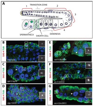
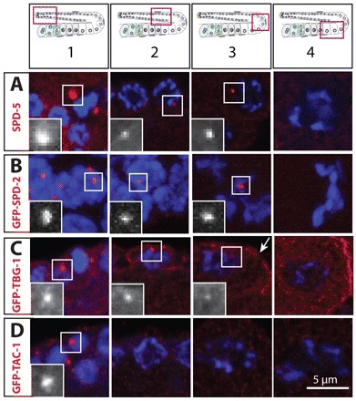
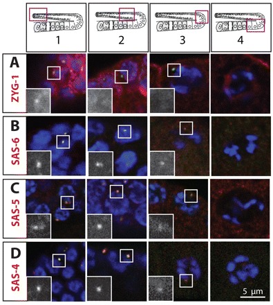
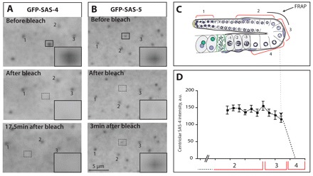
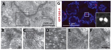
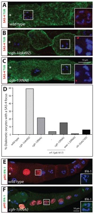
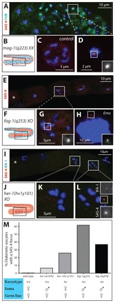
Centrosomes are the principal microtubule organizing centers (MTOCs) of animal cells and comprise a pair of centrioles surrounded by pericentriolar material (PCM). Centriole number must be carefully regulated, notably to ensure bipolar spindle formation and thus faithful chromosome segregation. In the germ line of most metazoan species, centrioles are maintained during spermatogenesis, but eliminated during oogenesis. Such differential behavior ensures that the appropriate number of centrioles is present in the newly fertilized zygote. Despite being a fundamental feature of sexual reproduction in metazoans, the mechanisms governing centriole elimination during oogenesis are poorly understood. Here, we investigate this question in C. elegans. Using antibodies directed against centriolar components and serial-section electron microscopy, we establish that centrioles are eliminated during the diplotene stage of the meiotic cell cycle. Moreover, we show that centriole elimination is delayed upon depletion of the helicase CGH-1. We also find that somatic cells make a minor contribution to this process, and demonstrate that the germ cell karyotype is important for timely centriole elimination. These findings set the stage for a mechanistic dissection of centriole elimination in a metazoan organism.


Figures

Fig. 1.
Fig. 1.
Requirements for centriolar proteins in the hermaphrodite gonad. (A) Schematic representation of the hermaphrodite gonad. For simplicity, the gonad is subdivided into four regions: (1) proliferating GCs; (2) GCs in pachytene; (3) GCs after pachytene; (4) GCs in diplotene after the loop. Note diakinesis oocytes (marked –1, –2, –3) prior to the spermatheca, with six condensed bivalent chromosomes. Sheath cell nuclei are depicted in blue and for simplicity only on one side of the gonad. (B-G) Region 1 from C. elegans gonads at the fourth larval stage (L4) of the indicated genotypes stained for α-tubulin (green), SAS-4 (red in the merged images and shown alone in magnified insets) and DNA (blue). Insets are magnified twofold. Note that the spd-2(oj29) GCN highlighted in C has two SAS-4 foci. Arrowheads point to clearly enlarged GCN.
Fig. 2.
Fig. 2.
Distribution of PCM proteins. Young adult hermaphrodite gonads stained for the indicated PCM components (red in the merged images and shown alone in magnified insets) and DNA (blue). Insets are magnified twofold. Schematic representations above the panels indicate positions of regions 1-4 in the gonad. The four panels of each row do not necessarily come from the same gonad.
Fig. 3.
Fig. 3.
Distribution of centriolar proteins. Young adult hermaphrodite gonads stained for the indicated centriolar proteins (red in the merged images and shown alone in magnified insets), IFA-1 (green) and DNA (blue). Insets are magnified twofold. Schematic representations above the panels indicate positions of regions 1-4 in the gonad. The four panels of each row do not necessarily come from the same gonad. Note that two foci are visible next to the GCN highlighted in C, region 1 and B, region 2.
Fig. 4.
Fig. 4.
Dynamics of centriolar proteins and kinetics of centriole elimination. (A,B) Images before (top), just after (middle) or at the indicated time after (bottom) photobleaching in region 2/3 of gonads expressing GFP-SAS-4 (A) or GFP-SAS-5 (B). 1, 2 and 3 mark reference centrioles in the field of view that were not bleached. Insets are magnified 3.5-fold. See also supplementary material Movies 1 and 2. (C) Schematic representation indicating the location of the FRAP experiments. (D) SAS-4 centriolar signal intensity in cells located in regions 2, 3 or 4, as indicated, expressed as percentage increase over the surrounding cytoplasmic signal (arbitrary units, a.u.). Dashed line between C and D indicates the approximate location of abrupt SAS-4 signal loss. Shown are mean and s.e.m., n=34.
Fig. 5.
Fig. 5.
Ultrastructural analysis and super-resolution microscopy of centrioles in pachytene. (A-F) TEM serial sections of centrioles in region 2; B,C,E and F are consecutive sections of the inset in A and show two disengaged mature centrioles (marked 1 and 2). D highlights nine microtubules in one centriole from the inset in C. (G) SIM of centrioles in region 2. Gonad from animal expressing GFP-SAS-4 stained for GFP (red in the merged images and shown alone in magnified inset) and DNA (blue).
Fig. 6.
Fig. 6.
CGH-1 promotes timely centriole elimination. (A-C) Region 4 from young adult hermaphrodites of the indicated genotypes stained for α-tubulin (green), SAS-4 (red in the merged images and shown with DNA in the insets) and DNA (blue). Note that the cgh-1(ok492) diakinesis GC highlighted in B is clearly cellularized. Note also that the inset for the DNA in these panels is a projection to visualize the DNA, whereas the low magnification image represents only one of the z-sections. The most mature GC/oocyte is to the left and marked by an asterisk. (D) Percentage of diakinesis GCs with SAS-4 foci. n and P-values (compared with wild type; Fisher’s exact test): wild type, n=79; cgh-1(ok492), n=22, P<0.0001; cgh-1(RNAi), n=93, P<0.0001; rrf-1(pk1417), n=27, P=0.2547; rrf-1(pk1417) cgh-1(RNAi), n=33, P=0.0018 and P=0.6120 compared with cgh-1(RNAi); wee-1.3(RNAi), n=100, P=1; cki-2(ok2105), n=67, P=0.042. (E,F) Wild-type (E) and cgh-1(RNAi) (F) gonads of young adult hermaphrodites stained for phospho-H3 (red), IFA-1 (green in the merged images and shown with DNA in insets) and DNA (blue). Note that in ∼16% of cgh-1(RNAi) phospho-H3-positive nuclei an IFA-1 focus was observed (n=67) whereas no such cases were observed in the wild type (n=35). Insets are magnified twofold.
Fig. 7.
Fig. 7.
The karyotype contributes to centriole elimination. (A,C,D) Region 3/4 from a mog-1(q233)XX animal (A,D) stained for α-tubulin (green), SAS-4 (red) and DNA (blue). D shows a high magnification view of the area denoted in A, with SAS-4 signal highlighted. A heterozygous diakinetic hermaphrodite oocyte is shown in panel C for comparison. (B) Schematic representation of a mog-1(q233)XX gonad in which hermaphroditic somatic cells (red) encase male GCs (blue). (E,G,H) Region 4 from a fog-1(q253)X0 animal (E,G,H) stained for SAS-4 (red) and DNA (blue). G shows a high magnification view of the area denoted in E, with SAS-4 signal highlighted. The most mature GC/oocyte is to the left and marked by an asterisk. (F) Schematic representation of a fog-1(q253)X0 gonad, in which male somatic cells (blue) encase female GCs (red). (H) Emo oocyte. The number of SAS-4 foci in fog-1(q253)X0 animals ranged from zero to eight, both in diakinesis and Emo oocytes, with the majority having no (32% for diakinesis, 72% for Emo) or one SAS-4 focus (31% for diakinesis, 9% for Emo). We interpret the occurrence of oocytes with more than two SAS-4 foci as centrioles breaking apart or as supernumerary centriole formation during endoreduplicating cycles. (I,K,L) Region 4 from a her-1(hv1y101)X0 animal stained for SAS-4 (red), IFA-1 (green) and DNA (blue). K and L show high magnification views of the areas denoted in I. The most mature GC/oocyte is to the left and marked by an asterisk. (J) Schematic representation of a her-1(hv1y101)X0 gonad in which female X0 GCs (red) are contained in a female gonad (red). (M) Karyotype, sex of the soma and the germ line, as well as percentage of diakinesis GCs with SAS-4 foci. n and P-values (compared with wild type; Fisher’s exact test): wild type, n=79; tra-1(e1076), n=47, P=0.050; fog-1(q253), n=73, P<0.0001; fog-3(q470), n=38, P<0.0001; her-1(hv1y101), n=163, P<0.0001. Insets are magnified twofold.

References

    1. Audhya A., Hyndman F., McLeod I. X., Maddox A. S., Yates J. R., 3rd, Desai A., Oegema K. (2005). A complex containing the Sm protein CAR-1 and the RNA helicase CGH-1 is required for embryonic cytokinesis in C. elegans. J. Cell Biol. 171, 267–279 - PMC - PubMed
    1. Baker R. E., Masison D. C. (1990). Isolation of the gene encoding the Saccharomyces cerevisiae centromere-binding protein CP1. Mol. Cell. Biol. 10, 2458–2467 - PMC - PubMed
    1. Barton M. K., Kimble J. (1990). fog-1, a regulatory gene required for specification of spermatogenesis in the germ line of C. elegans. Genetics 125, 29–39 - PMC - PubMed
    1. Barton M. K., Schedl T. B., Kimble J. (1987). Gain-of-function mutations of fem-3, a sex-determination gene in C. elegans. Genetics 115, 107–119 - PMC - PubMed
    1. Bean C. J., Schaner C. E., Kelly W. G. (2004). Meiotic pairing and imprinted X chromatin assembly in C. elegans. Nat. Genet. 36, 100–105 - PMC - PubMed

Publication types