. 2012 Jun;78(12):4400-11.
doi: 10.1128/AEM.07895-11. Epub 2012 Apr 6.

Functional specificity of extracellular nucleases of Shewanella oneidensis MR-1

Affiliations

Functional specificity of extracellular nucleases of Shewanella oneidensis MR-1

Magnus Heun et al. Appl Environ Microbiol. 2012 Jun.

Abstract

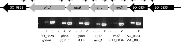
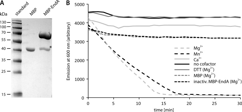
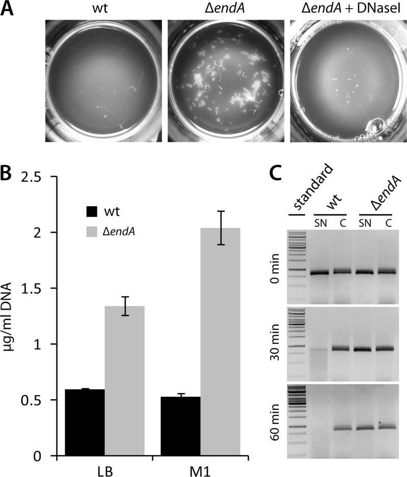
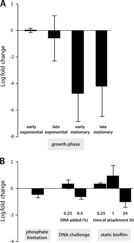
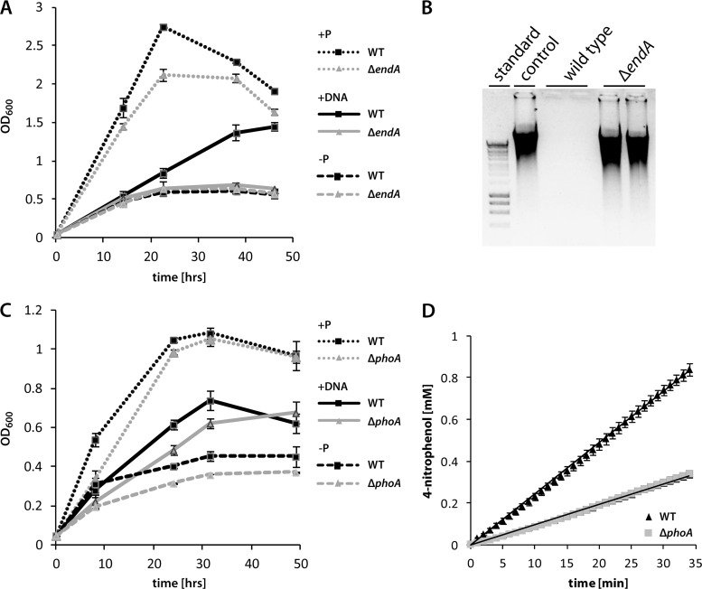
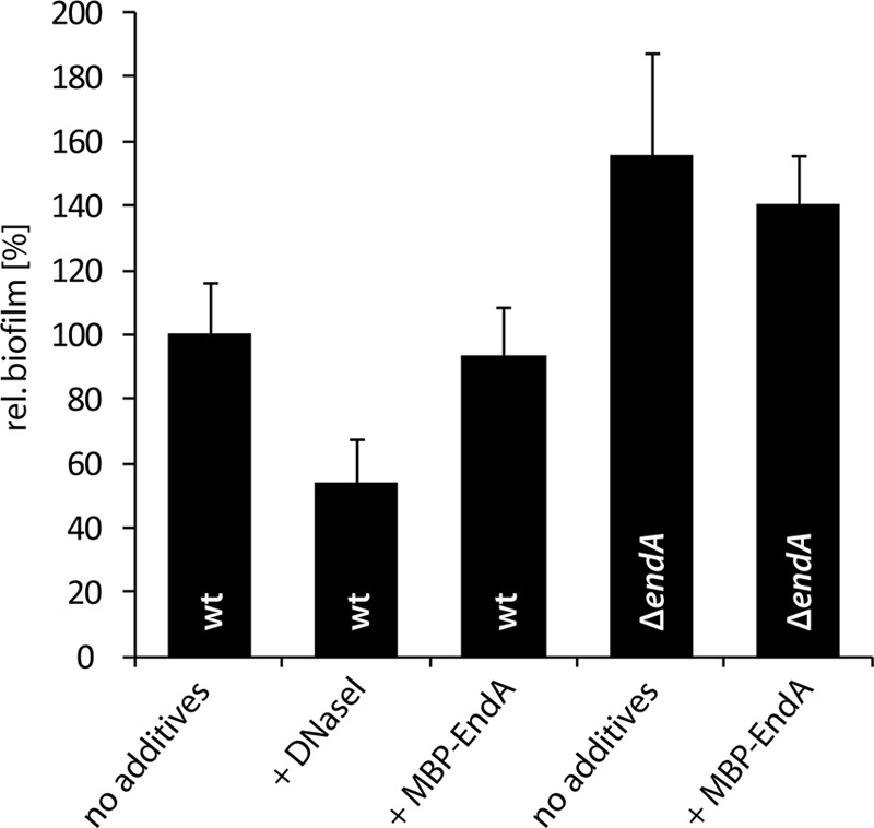
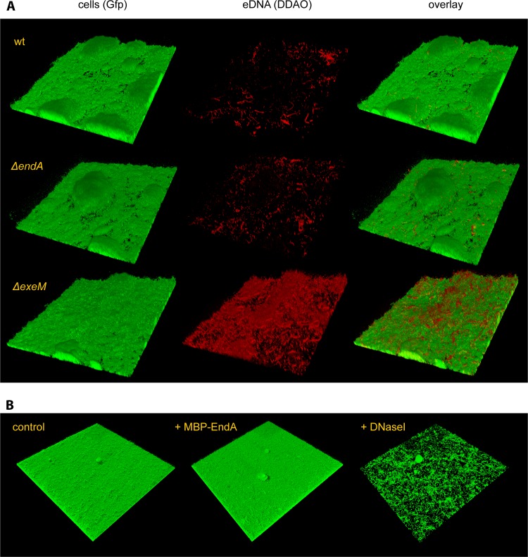
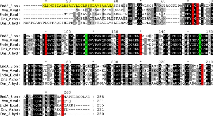
Bacterial species such as Shewanella oneidensis MR-1 require extracellular nucleolytic activity for the utilization of extracellular DNA (eDNA) as a source of nutrients and for the turnover of eDNA as a structural matrix component during biofilm formation. We have previously characterized two extracellular nucleases of S. oneidensis MR-1, ExeM and ExeS. Although both are involved in biofilm formation, they are not specifically required for the utilization of eDNA as a nutrient. Here we identified and characterized EndA, a third extracellular nuclease of Shewanella. The heterologously overproduced and purified protein was highly active and rapidly degraded linear and supercoiled DNAs of various origins. Divalent metal ions (Mg(2+) or Mn(2+)) were required for function. endA is cotranscribed with phoA, an extracellular phosphatase, and is not upregulated upon phosphostarvation. Deletion of endA abolished both extracellular degradation of DNA by S. oneidensis MR-1 and the ability to use eDNA as a sole source of phosphorus. PhoA is not strictly required for the exploitation of eDNA as a nutrient. The activity of EndA prevents the formation of large cell aggregates during planktonic growth. However, in contrast to the findings for ExeM, endA deletion had only minor effects on biofilm formation. The findings strongly suggest that the extracellular nucleases of S. oneidensis exert specific functions required under different conditions.


Figures

Fig 1
Fig 1
Genetic and transcriptomic organization of endA and phoA. (Top) Schematic representation of the genetic organization. Gene numbers or corresponding annotations are given. Arrows indicate the positions of the corresponding primer pairs. (Bottom) RT analysis of the transcript. −, negative control with no reverse transcriptase added to the reaction mixture; +, standard RT-PCR; c, control reaction with chromosomal DNA as the template. CHP, conserved hypothetical protein.
Fig 2
Fig 2
Purification and in vitro activity of MPB-EndA. (A) Polyacrylamide gel electrophoresis of proteins after enrichment using amylose resin. Unfused MBP was produced and enriched in parallel (center lane). MBP-EndA migrated at a position corresponding to a molecular mass of about 70 kDa, in close agreement with the estimated mass of 73.1 kDa. A single major contaminating band (marked by an asterisk), likely representing MBP (42 kDa), was visible after enrichment. (B) Activity of highly enriched MBP-EndA on the purified vector pBluescript. The activity was determined by the loss of fluorescence of DNA-bound GelRed nucleic acid stain due to DNA degradation. Several cofactors (Mg2+, Mn2+, Ca2+) were tested, and control assays were carried out either with no cofactor, after the addition of DTT, after heat inactivation of MBP-EndA, or with MBP that was analogously produced and purified. Only the addition of Mg2+ or Mn2+ as a cofactor to EndA resulted in rapid degradation of DNA.
Fig 3
Fig 3
In vivo activity of EndA in medium supernatants. (A) Flocculation phenotype of ΔendA mutants. Displayed are top views of cultures of the indicated strains grown for 72 h in M1 mineral medium. ΔendA mutants form aggregates (center) that are readily dispersed upon addition of DNase I (right). Heat-inactivated DNase I had no effect. Similar flocculation also occurred in complex LB medium. (B) Amounts of eDNA in medium supernatants of wild-type (filled bars) and ΔendA mutant (shaded bars) cultures grown in LB (left) or M1 (right) medium. Error bars, standard deviations. (C) Nuclease activities in medium supernatants of the wild type and the ΔendA mutant. The cultures were grown to exponential phase in LB medium, and production of new protein was blocked by the addition of chloramphenicol. Subsequently, an 833-bp DNA fragment was added directly to the supernatants of the cultures (SN) and to the supernatants of cultures in which the cell sediment had been washed prior to the addition of the PCR fragment (C). After the indicated times, aliquots were removed and analyzed by agarose gel electrophoresis. DNA degradation was observed only in the supernatants of unwashed wild-type cultures, strongly indicating that EndA is the main nuclease in the supernatant and likely is not associated with the cell envelope.
Fig 4
Fig 4
Regulation of endA. endA transcript levels were determined by qRT-PCR. (A) Growth-dependent regulation of endA. Displayed is the regulation of cells in the late-exponential-growth phase (OD600, 2.0) and in the early- and late-stationary-growth phases (OD600, 4.0 and 6.0) compared to that in the early-exponential-growth phase in LB medium (OD600, 0.6). (B) Regulation of endA in response to different environmental stimuli. (Left) Regulation during growth on DNA compared to growth on NaH2PO4; (center) regulation of endA 30 min after the addition of sublethal amounts of eDNA to exponentially growing cultures compared to nonchallenged cultures; (right) differential regulation of endA in planktonic cells and surface-associated cells harvested from static biofilm cultures at the indicated time points. Error bars, standard deviations.
Fig 5
Fig 5
Contributions of EndA and PhoA to aerobic growth on eDNA as the sole source of phosphate. (A) Contribution of EndA to growth on eDNA. The growth of the wild type (filled squares) and the ΔendA mutant (shaded triangles) was followed for 62 h in M1 mineral medium supplemented with either 0.86 mM NaH2PO4 (dotted lines), salmon sperm DNA (0.5 g · liter−1) (solid lines), or no source of phosphorus (dashed lines). Error bars, standard deviations. (B) Degradation of eDNA during growth. Displayed is an agarose gel separation of added salmon sperm DNA after 18 h of incubation in plain M1 medium (control), with the wild type, and with a ΔendA mutant. In the absence of EndA, little DNA degradation occurred. (C) Contribution of PhoA to growth on eDNA as a sole source of phosphorus. The growth of the wild type (filled squares) and the ΔphoA mutant (shaded triangles) was followed for 62 h in M1 mineral medium supplemented with either 0.86 mM NaH2PO4 (dotted lines), salmon sperm DNA (0.5 g · liter−1) (solid lines), or no source of phosphorus (dashed lines). The utilization of eDNA as a source of phosphorus was not significantly affected in the ΔphoA mutant. (D) Activity of PhoA in medium supernatants. Phosphatase activity in M1 medium supernatants was determined for wild-type (filled triangles) and ΔphoA mutant (shaded squares) cultures. The graph displays the formation of 4-nitrophenol from p-nitrophenyl phosphate over time. Supernatants of wild-type cultures exhibit significantly higher phosphatase activity than supernatants of ΔphoA mutant cultures. Error bars, standard deviations.
Fig 6
Fig 6
Role of EndA in biofilm formation under static conditions. Wild-type and ΔendA biofilms were grown in microtiter plates for 24 h, and DNase I or MBP-EndA was added as indicated. The values are means of three replicates. Error bars, standard deviations. rel., relative.
Fig 7
Fig 7
Role of EndA in biofilm formation under hydrodynamic conditions. (A) (Left) The formation of biofilms by Gfp-tagged wild-type and ΔendA and ΔexeM nuclease mutant strains under hydrodynamic conditions was monitored by CLSM after 48 h of incubation. (Center) eDNA was then visualized by DDAO staining. (Right) The corresponding overlays are displayed. (B) A wild-type biofilm formed after 24 h in flow chambers was treated with MBP-EndA (center) or DNase I (right) for 45 min. The lateral edge of each micrograph measures 250 μm.
Fig 8
Fig 8
Sequence alignments of S. oneidensis MR-1 EndA with known nucleases. endA_S.on, S. oneidensis MR-1 EndA; Vvn_V.vul, V. vulnificus Vvn; endA_E.col, E. coli EndA; Dns_V.cho, V. cholerae Dns; Dns_A.hyd, A. hydrophila Dns. Areas of significant similarity are highlighted in gray and black. The gradient indicates the degree of conservation, from black (fully conserved) to light grey (less well conserved). The cysteine residues required for disulfide bond formation are highlighted in red, and residues involved in coordinating the metal cofactor are highlighted in green. The putative signal sequence of EndA is highlighted in yellow. The amino acid numbering of the longest peptide (Dns_A.hyd) is used, and every 10th position is indicated by either a number or an asterisk.

References

    1. Aiba H, Adhya S, de Crombrugghe B. 1981. Evidence for two functional gal promoters in intact Escherichia coli cells. J. Biol. Chem. 256:11905–11910 - PubMed
    1. Altermark B, Smalas AO, Willassen NP, Helland R. 2006. The structure of Vibrio cholerae extracellular endonuclease I reveals the presence of a buried chloride ion. Acta Crystallogr. D Biol. Crystallogr. 62:1387–1391 - PubMed
    1. Baldwin DS. 1998. Reactive “organic” phosphorus revisited. Water Res. 32:2265–2270
    1. Bendtsen JD, Nielsen H, von Heijne G, Brunak S. 2004. Improved prediction of signal peptides: SignalP 3.0. J. Mol. Biol. 340:783–795 - PubMed
    1. Berne C, Kysela DT, Brun YV. 2010. A bacterial extracellular DNA inhibits settling of motile progeny cells within a biofilm. Mol. Microbiol. 77:815–829 - PMC - PubMed

Publication types

LinkOut - more resources