

Baseline cardiovascular risk predicts subsequent changes in resting brain function
- PMID: 22492519
- PMCID: PMC3361601
- DOI: 10.1161/STROKEAHA.111.638437
Baseline cardiovascular risk predicts subsequent changes in resting brain function
Abstract
Background and purpose: The Framingham Heart Study group cardiovascular disease risk profile (FCRP) score was used to assess the relationship between baseline cardiovascular risk and subsequent changes in resting state cerebral blood flow (CBF) in cognitively normal older participants from the Baltimore Longitudinal Study of Aging.
Methods: Ninty-seven cognitively normal participants underwent annual resting-state positron emission tomography scans at baseline and over a period of up to 8 years (mean interval, 7.4 years). Images quantifying voxel-wise longitudinal rates of CBF change were calculated and used to examine the relationship between baseline FCRP score and changes over time in regional CBF. Individual components of the FCRP score (age, cholesterol, blood pressure, smoking status, and type 2 diabetes) were also correlated with changes in regional CBF to examine the independent contributions of each component to the overall pattern of change.
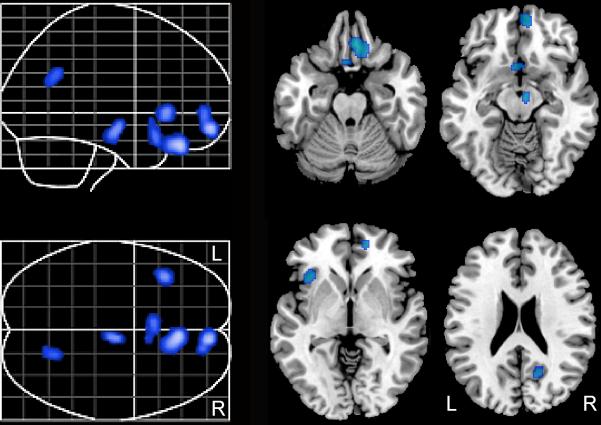
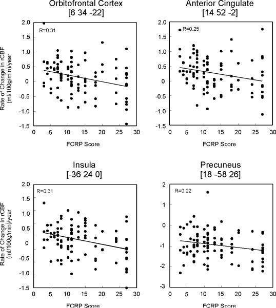
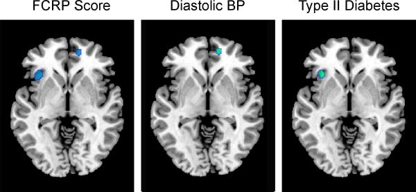
Results: Higher baseline FCRP scores were associated with accelerated longitudinal decline in CBF in orbitofrontal, medial frontal/anterior cingulate, insular, precuneus, and brain stem regions. Of the components that comprise the FCRP score, higher diastolic blood pressure and diabetes were associated independently with greater decline in the medial frontal/anterior cingulate and insular regions, respectively.
Conclusions: Baseline cardiovascular risk factors are associated with greater rates of decline in resting state regional brain function. The regions showing accelerated decline participate in higher-order cognitive processes and are also vulnerable to age-related neuropathology. These results, in conjunction with other studies, encourage early treatment of cardiovascular risk factors in older individuals.
Figures
Similar articles
-
Changes in brain function occur years before the onset of cognitive impairment.J Neurosci. 2013 Nov 13;33(46):18008-14. doi: 10.1523/JNEUROSCI.1402-13.2013. J Neurosci. 2013. PMID: 24227712 Free PMC article.
-
Patterns of regional cerebral blood flow associated with low hemoglobin in the Baltimore Longitudinal Study of Aging.J Gerontol A Biol Sci Med Sci. 2012 Sep;67(9):963-9. doi: 10.1093/gerona/gls121. Epub 2012 May 2. J Gerontol A Biol Sci Med Sci. 2012. PMID: 22552368 Free PMC article.
-
Framingham cardiovascular risk profile correlates with impaired hippocampal and cortical vasoreactivity to hypercapnia.J Cereb Blood Flow Metab. 2011 Feb;31(2):671-9. doi: 10.1038/jcbfm.2010.145. Epub 2010 Sep 15. J Cereb Blood Flow Metab. 2011. PMID: 20842159 Free PMC article.
-
Elevated Markers of Inflammation Are Associated With Longitudinal Changes in Brain Function in Older Adults.J Gerontol A Biol Sci Med Sci. 2018 May 9;73(6):770-778. doi: 10.1093/gerona/glx199. J Gerontol A Biol Sci Med Sci. 2018. PMID: 29304217 Free PMC article.
-
Lower cardiac index levels relate to lower cerebral blood flow in older adults.Neurology. 2017 Dec 5;89(23):2327-2334. doi: 10.1212/WNL.0000000000004707. Epub 2017 Nov 8. Neurology. 2017. PMID: 29117962 Free PMC article.
Cited by
-
Cerebrovascular complications of diabetes: focus on cognitive dysfunction.Clin Sci (Lond). 2016 Oct 1;130(20):1807-22. doi: 10.1042/CS20160397. Clin Sci (Lond). 2016. PMID: 27634842 Free PMC article. Review.
-
The Effects of Aerobic Exercise on Cognitive and Neural Decline in Aging and Cardiovascular Disease.Curr Geriatr Rep. 2014 Dec;3(4):282-290. doi: 10.1007/s13670-014-0101-x. Curr Geriatr Rep. 2014. PMID: 25750853 Free PMC article.
-
Compensatory functional reorganization may precede hypertension-related brain damage and cognitive decline: a functional magnetic resonance imaging study.J Hypertens. 2017 Jun;35(6):1252-1262. doi: 10.1097/HJH.0000000000001293. J Hypertens. 2017. PMID: 28169883 Free PMC article.
-
Dynamic association between perfusion and white matter integrity across time since injury in Veterans with history of TBI.Neuroimage Clin. 2016 Dec 23;14:308-315. doi: 10.1016/j.nicl.2016.12.017. eCollection 2017. Neuroimage Clin. 2016. PMID: 28210542 Free PMC article.
-
Volumetric brain MRI signatures of heart failure with preserved ejection fraction in the setting of dementia.Magn Reson Imaging. 2024 Jun;109:49-55. doi: 10.1016/j.mri.2024.02.016. Epub 2024 Feb 29. Magn Reson Imaging. 2024. PMID: 38430976 Free PMC article.
References
-
- Waldstein SR, Wendell CR. Neurocognitive function and cardiovascular disease. J Alzheimers Dis. 2010;20:833–842. - PubMed
-
- de Toledo Ferraz Alves TC, Ferreira LK, Wajngarten M, Busatto GF. Cardiac disorders as risk factors for alzheimer's disease. J Alzheimers Dis. 2010;20:749–763. - PubMed
-
- Sakurai H, Hanyu H, Sato T, Kanetaka H, Shimizu S, Hirao K, et al. Vascular risk factors and progression in alzheimer's disease. Geriatr Gerontol Int. 2011;11:211–214. - PubMed
Publication types
MeSH terms
Grants and funding
LinkOut - more resources
Full Text Sources
Medical
