. 2012 Jun;61(6):1423-33.
doi: 10.2337/db11-0961. Epub 2012 Apr 9.

Cellular responses to the metal-binding properties of metformin

Affiliations

Cellular responses to the metal-binding properties of metformin

Lisa Logie et al. Diabetes. 2012 Jun.

Abstract

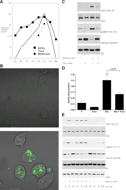
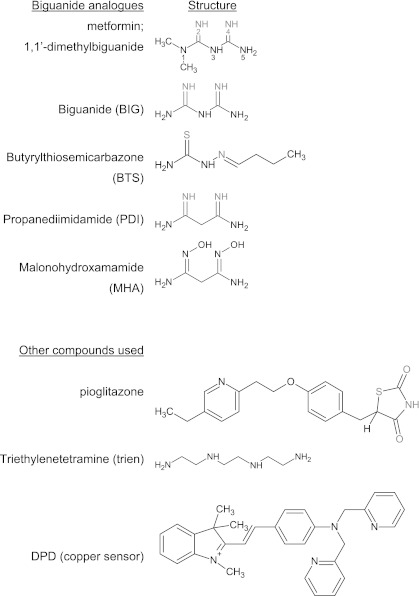
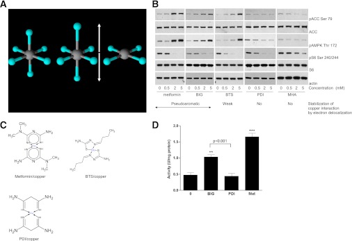
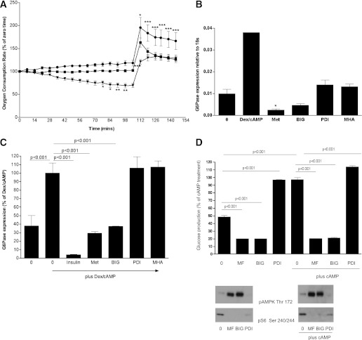
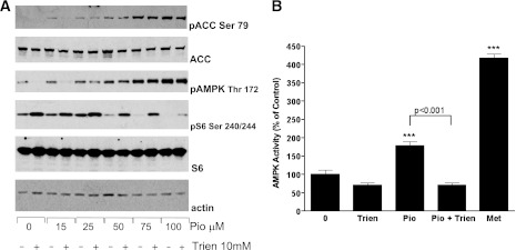
In recent decades, the antihyperglycemic biguanide metformin has been used extensively in the treatment of type 2 diabetes, despite continuing uncertainty over its direct target. In this article, using two independent approaches, we demonstrate that cellular actions of metformin are disrupted by interference with its metal-binding properties, which have been known for over a century but little studied by biologists. We demonstrate that copper sequestration opposes known actions of metformin not only on AMP-activated protein kinase (AMPK)-dependent signaling, but also on S6 protein phosphorylation. Biguanide/metal interactions are stabilized by extensive π-electron delocalization and by investigating analogs of metformin; we provide evidence that this intrinsic property enables biguanides to regulate AMPK, glucose production, gluconeogenic gene expression, mitochondrial respiration, and mitochondrial copper binding. In contrast, regulation of S6 phosphorylation is prevented only by direct modification of the metal-liganding groups of the biguanide structure, supporting recent data that AMPK and S6 phosphorylation are regulated independently by biguanides. Additional studies with pioglitazone suggest that mitochondrial copper is targeted by both of these clinically important drugs. Together, these results suggest that cellular effects of biguanides depend on their metal-binding properties. This link may illuminate a better understanding of the molecular mechanisms enabling antihyperglycemic drug action.


Figures

FIG. 1.
FIG. 1.
Trien opposes the action of metformin on AMPK and signaling to S6. A: Graph of the available stability constants for EDTA, triethylaminetetramine (trien), and the cumulative constant for a 2:1 dimethylbiguanide (metformin)/metal complex (data obtained from Refs. 17,25,26). Biguanides do not form stable complexes with zinc or alkaline earth metals (17). B: Cells placed in serum-free medium treated with (bottom)/without (top) 10 mM trien for 18 h were treated with the copper-binding probe followed by washout and image capture on the confocal microscope. Scale bar, 10 μm. C: Preincubation of cells with trien inhibits metformin’s effects on AMPK Thr172 phosphorylation (pAMPK), ACC Ser79 phosphorylation (pACC), and S6 Ser240/244 phosphorylation (pS6). H4IIE cells were grown in serum-free medium for 18 h in the presence or absence of 10 mM trien, followed by stimulation with or without 2 mM metformin. Lysates were prepared as described in research design and methods then subjected to SDS-PAGE, followed by immunoblotting with the antibodies indicated. D: Cells were treated as in C, except 1 mM metformin (Met) was used, before lysis, immunoprecipitation, and AMPK assay as described in research design and methods. Statistical significance was determined by one-way ANOVA followed by Tukey post hoc test, ***P < 0.001 with respect to control. The significance of other column-to-column differences are presented next to a horizontal line identifying the two columns. Errors are SEM. E: Cells were treated as in C but in the presence of a dose response of trien. (A high-quality digital representation of this figure is available in the online issue.)
FIG. 2.
FIG. 2.
Compounds used in this study. Known metal coordinating ligands appear for each biguanide analog in gray. In pioglitazone, gray highlighting denotes the region of similarity between glitazones and metformin, along with the sulfur atom that undergoes oxidation-enabling linearization of the thiazolidinedione moiety. Chemical structures were drawn using ChemSketch (Advanced Chemistry Development, Inc.).
FIG. 3.
FIG. 3.
Interruption of π-electron delocalization intrinsic to biguanides results in compounds that selectively target S6 phosphorylation, lacking effects on AMPK. A: Commonly, distortion of copper d9 octahedral coordination geometry (left) results in elongated axial ligands (middle). The extreme case of axial ligand elongation is the square planar structure (right). Copper is induced to adopt this geometry when metformin binds. B and C: H4IIE cells were grown in serum-free medium for 2 h, followed by stimulation with or without the metformin analogs shown, at the concentrations shown. Lysates were prepared for immunoblotting with the antibodies indicated. The chemical structures of metformin, BTS, and PDI complexed with divalent copper are shown in C. Metformin and BIG form pseudoaromatic planar complexes with copper in a square planar geometry, characterized by fairly uniform intermediate bond lengths in the ring, stabilized by π-electron delocalization (–14). Thiosemicarbazones such as BTS also form extensively conjugated ring structures with copper (33); however, data on bond lengths (30,31), supported by spectroscopic studies (42), indicate that the resonance form shown predominates, with reduced delocalization of electron density and significant deviation from planarity compared with biguanides (11,14,31). PDI has π-electron delocalization in the ring abolished by substitution of N3 (12,32) and is not planar. D: H4IIE cells that had been deprived of serum for 2 h were treated with 2 mM of the agents shown for 3 h (metformin [Met]), before AMPK assay as described in research design and methods. Statistical analysis was carried out as in Fig. 1D. **P < 0.01, ***P < 0.001. (A high-quality digital representation of this figure is available in the online issue.)
FIG. 4.
FIG. 4.
Effect of biguanides and PDI on mitochondrial respiration, gluconeogenesis, and glucose production. A: H4IIE cells were grown in serum-free medium overnight followed by stimulation without (squares) or with metformin (inverted triangles) or PDI (circles) as indicated. Cells were treated with drug 1 h before measurements started. Using a Seahorse analyzer (Seahorse Bioscience), OCR was measured. At 105 min, 100 μM 2,4 dinitrophenol was added to uncouple the electron transport chain from control by ATP synthesis in order to reveal the maximal respiration rate. There were no statistically significant differences in OCR between different treatment groups at time zero. Data are normalized to 100 at the start of the experiment. Statistical analysis was carried out as in Fig. 1D, except that a two-way ANOVA was performed. B and C: H4IIE cells were grown for 3 days, then serum-starved overnight, followed by stimulation for 8 h with or without 500 nM dexamethasone (Dex)/100 μM CPT-cAMP, coincubated with 10 nM insulin or the metformin analogs shown (2 mM of each, abbreviations are as in the main text). Expression of G6Pase was measured by RT-PCR, using conditions described in research design and methods. Statistical analysis was carried out as in Fig. 1D. D: Primary hepatocytes were treated with or without 2 mM metformin (MF), 5 mM BIG, or 5 mM PDI for 12 h with or without 100 μM Bt2-cAMP and glucose production was measured as described in research design and methods. The cells were lysed and effects of the drugs on cell signaling verified by Western blotting (bottom). Statistical analysis was carried out as in Fig. 1D. *P < 0.05, **P < 0.01, ***P < 0.001.
FIG. 5.
FIG. 5.
Effect of metformin analogs on the copper-specific fluorescent probe. Cells were serum-starved for 2 h before treatment for 3 h without (A) or with the biguanides metformin (left) and BIG (right) (B) or PDI (left) and MHA (right) (C). In each case, the cells were incubated in the presence of the copper-specific probe, before washout and visualization on the confocal microscope as discussed in research design and methods. Scale bars, 10 μm. (A high-quality digital representation of this figure is available in the online issue.)
FIG. 6.
FIG. 6.
Effect of trien on cell signaling responses to pioglitazone. A: H4IIE cells were grown in serum-free medium for 18 h in the presence or absence of 10 mM trien, followed by stimulation with or without differing doses of pioglitazone, as indicated. Lysates were prepared for immunoblotting with the antibodies indicated as described in research design and methods. B: H4IIE cells were grown in serum-free medium for 18 h in the absence or presence of 10 mM trien. Individual dishes were then stimulated without or with 15 μM pioglitazone (Pio) for 3 h. Total of 2 mM metformin (Met) was added to further dishes for 3 h as a positive control, and then the samples were prepared for AMPK assay as described in research design and methods. Statistical analysis was carried out as in Fig. 1D. ***P < 0.001.
FIG. 7.
FIG. 7.
Evidence that metformin and pioglitazone regulate mitochondrial copper. A: H4IIE cells were grown in serum-free medium for 2 h before treatment with (right) or without (left) 100 μM pioglitazone for 3 h. In each case, the cells were incubated in the presence of the copper-specific probe, before washout and visualization on the confocal microscope as discussed in research design and methods. Scale bars, 10 μm. B and C: Cells were serum-starved for 2 h before treatment for 3 h with 100 μM pioglitazone (B) or 2 mM metformin (C). The cells were incubated in the presence of the copper-specific probe (green channel, ii) and a mitochondrial marker (far red channel, iv), before washout and visualization on the confocal microscope as discussed in research design and methods. Hoechst staining (blue channel, i) marks the nucleus. All channels are merged in v to demonstrate colocalization. Panel iii is phase-contrast. (A high-quality digital representation of this figure is available in the online issue.)

References

    1. Slotta KH, Tschesche R. Über Biguanide, II.: Die blutzucker-senkende Wirkung der Biguanide. Ber Dtsch Chem Ges 1929;62:1398–1405
    1. Hawgood BJ. Karl Heinrich Slotta (1895–1987) biochemist: snakes, pregnancy and coffee. Toxicon 2001;39:1277–1282 - PubMed
    1. Shapiro SL, Parrino VA, Freedman L. Hypoglycemic agents III. N-Alkyl- and arylalkylbiguanides. J Am Chem Soc 1959;81:3728–3736
    1. Owen MR, Doran E, Halestrap AP. Evidence that metformin exerts its anti-diabetic effects through inhibition of complex 1 of the mitochondrial respiratory chain. Biochem J 2000;348:607–614 - PMC - PubMed
    1. El-Mir M-Y, Nogueira V, Fontaine E, Avéret N, Rigoulet M, Leverve X. Dimethylbiguanide inhibits cell respiration via an indirect effect targeted on the respiratory chain complex I. J Biol Chem 2000;275:223–228 - PubMed

Publication types