. 2012 Jun;135(Pt 6):1964-80.
doi: 10.1093/brain/aws075. Epub 2012 Apr 3.

Essential role of interleukin-6 in post-stroke angiogenesis

Affiliations

Essential role of interleukin-6 in post-stroke angiogenesis

Karen Gertz et al. Brain. 2012 Jun.

Abstract

Ambivalent effects of interleukin-6 on the pathogenesis of ischaemic stroke have been reported. However, to date, the long-term actions of interleukin-6 after stroke have not been investigated. Here, we subjected interleukin-6 knockout (IL-6(-/-)) and wild-type control mice to mild brain ischaemia by 30-min filamentous middle cerebral artery occlusion/reperfusion. While ischaemic tissue damage was comparable at early time points, IL-6(-/-) mice showed significantly increased chronic lesion volumes as well as worse long-term functional outcome. In particular, IL-6(-/-) mice displayed an impaired angiogenic response to brain ischaemia with reduced numbers of newly generated endothelial cells and decreased density of perfused microvessels along with lower absolute regional cerebral blood flow and reduced vessel responsivity in ischaemic striatum at 4 weeks. Similarly, the early genomic activation of angiogenesis-related gene networks was strongly reduced and the ischaemia-induced signal transducer and activator of transcription 3 activation observed in wild-type mice was almost absent in IL-6(-/-) mice. In addition, systemic neoangiogenesis was impaired in IL-6(-/-) mice. Transplantation of interleukin-6 competent bone marrow into IL-6(-/-) mice (IL-6(chi)) did not rescue interleukin-6 messenger RNA expression or the early transcriptional activation of angiogenesis after stroke. Accordingly, chronic stroke outcome in IL-6(chi) mice recapitulated the major effects of interleukin-6 deficiency on post-stroke regeneration with significantly enhanced lesion volumes and reduced vessel densities. Additional in vitro experiments yielded complementary evidence, which showed that after stroke resident brain cells serve as the major source of interleukin-6 in a self-amplifying network. Treatment of primary cortical neurons, mixed glial cultures or immortalized brain endothelia with interleukin 6-induced robust interleukin-6 messenger RNA transcription in each case, whereas oxygen-glucose deprivation did not. However, oxygen-glucose deprivation of organotypic brain slices resulted in strong upregulation of interleukin-6 messenger RNA along with increased transcription of key angiogenesis-associated genes. In conclusion, interleukin-6 produced locally by resident brain cells promotes post-stroke angiogenesis and thereby affords long-term histological and functional protection.


Figures
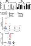
Figure 1
Figure 1
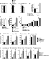
Early upregulation of angiogenesis-related genes after mild brain ischaemia is attenuated in IL-6−/− mice. IL-6 messenger RNA (A) and protein expression levels (B) were measured in ischaemic brain of wild-type mice after 30 min MCAO/reperfusion. (C and D) Transcription of IL-6 receptor (C) and gp130 (D) messenger RNA was assessed in IL-6−/− mice and wild-type littermate controls. Note that IL-6 receptor and, to a lesser extent gp130, showed prolonged upregulation after transient brain ischaemia. Relative messenger RNA expression is reported as the value normalized to tripeptidyl peptidase 2 (Tpp2) for each sample. n = 3–6 animals per group. *P < 0.05 versus sham. (E and F) IL-6−/− mice and wild-type littermate controls were exposed to 30 min MCAO/reperfusion or sham operation. Animals were euthanized at 2 and 10 days after brain ischaemia. Unpooled samples of ipsilateral brain tissue were processed for microarray analysis. Messenger RNA expression of 162 angiogenesis-associated genes (for detailed summary of gene expression see Supplementary Table 1) was analysed. (E) Ratio of gene expression of angiogenesis-associated genes in MCAO versus sham-operated animals. Note that at 2 days after 30 min MCAO/reperfusion, angiogenesis-associated genes as a class show significantly stronger activation in wild-type relative to IL-6−/− mice. (F) Significant increase in the ratio of gene expression in wild-type to IL-6−/− mice at 2 days after MCAO/reperfusion. Note that the expression of angiogenesis-related genes is comparable in IL-6−/− mice and wild-type controls both in the sham condition and at 10 days after transient brain ischaemia (ratio ∼1). Genes showing the strongest upregulation or the strongest downregulation are given in red and blue, respectively. Additionally, candidate genes VEGF receptor 2 (Kdr) and endothelial NO synthase (Nos3) confirmed by quantitative polymerase chain reaction (Supplementary Fig. 2H and I) are given (green triangles). n = 24 animals in total (n = 3 animals per intervention per genotype per time point). (E) and (F) represent the median, 10th, 25th, 75th and 90th percentiles as vertical boxes with error bars. One-way ANOVA on ranks followed by Newman–Keuls post hoc test. *P < 0.05. Adamts1 = a disintegrin-like and metallopeptidase; Anxa2 = Annexin A2; Bai1 = brain-specific angiogenesis inhibitor 1; Baiap2 = brain-specific angiogenesis inhibitor 1-associated protein; Bai3 = brain-specific angiogenesis inhibitor 3; Cx3cl1 = chemokine C-X3-C ligand 1; Cxcl4 chemokine = C-X-C ligand 4; Cx3cr1 = chemokine C-X3-C receptor 1; Cyr61 = cystein rich protein 61; Edn1 = Endothelin 1; Eng = Endoglin; Kdr = VEGF receptor 2; Nos3 = endothelial NO synthase; Ntrk2 = neurotrophic tyrosine kinase receptor typ2; Pdpn = Podoplanin; Plxnd1 = Plexin D1; Sema5a = Semaphorin 5a; Thbs1 = Thrombospondin 1.
Figure 2
Figure 2
Transplantation of IL-6 competent bone marrow into IL-6−/− mice does not rescue the angiogenic response to transient mild brain ischaemia. (A–N) Wild-type mice and IL-6−/− mice were lethally irradiated and then transplanted with bone marrow-derived from ubiquitously ‘green’ mice [TgN(beta-act-EGFP)1Osb]. All analyses were performed at 48 h after stroke. (A and B) The number of bone marrow-derived cells in the ischaemic striatum did not differ significantly between chimeric WTchi (WTGFP → IL-6+/+) and IL-6chi mice (WTGFP → IL-6−/−). n = 3 animals per group. (C) Despite engraftment of IL-6 competent bone marrow-derived cells in the ischaemic lesion, we did not detect IL-6 messenger RNA in the brains of IL-6chi mice. In contrast, MCAO elicited a strong increase in IL-6 messenger RNA transcription in the ischaemic lesion in WTchi mice. (D) Detection of 174 bp product of IL-6 complementary DNA by gel electrophoresis shows no amplification of an IL-6 specific product in IL-6chi mice. The minus-reverse transcriptase control is shown in lane 5. (E and F) IL-6 messenger RNA expression in spleen (E) and liver (F). (G) IL-6 protein levels in serum. n = 2–5 animals per group. (H–N) Expression of key angiogenesis-associated genes in the ischaemic hemisphere. n = 4–5 animals per group. (O–U) All analyses were performed at 28 days after stroke. (O and P) The number of bone marrow-derived cells in the ischaemic striatum differed significantly between WTchi and IL-6chi mice. n = 8–9 animals per group. (Q) Despite engraftment of IL-6 competent bone marrow-derived cells in the ischaemic lesion, we did not detect any IL-6 messenger RNA in the brains of IL-6chi mice. (R) Detection of 174 bp product of IL-6 complementary DNA by gel electrophoresis again shows no amplification of an IL-6 specific product in the brain of IL-6chi mice. (S) In contrast, IL-6 messenger RNA expression in spleen (left) and liver (right) is clearly detectable in IL-6chi mice. Minus-reverse transcriptase controls are shown in lane 5 (R) as well as in lanes 5 and 10 (S). (T) IL-6 protein levels in serum. n = 3–6 animals per group. (U) The density of perfused microvessels was determined using endovascular, auto-fluorescent Evans blue staining and tiled-field imaging. Note significant inducing effect of ischaemia on vessel density in WTchi mice, which was significantly attenuated in IL-6chi mice. n = 5–6 animals per group. *P < 0.05 relative to sham, #P < 0.05 between recipient genotypes. Scale bar: B = 100 μm; P = 38 μm.
Figure 3
Figure 3
Characterization of the cellular source of IL-6 in brain tissue. Primary cortical neurons, mixed glial cultures and immortalized brain endothelia were subjected to oxygen–glucose deprivation as described in the text (A–C). Oxygen–glucose deprivation did not lead to increased IL-6 gene transcription in isolated cultures. Cultures were exposed to recombinant IL-6. Stimulation of primary cortical neurons (D), mixed glial cultures (E) and of immortalized brain endothelia (F) resulted in significantly increased IL-6 messenger RNA transcription. (G) Stimulation of bEnd.3 cells by exposure to IL-6 resulted in significantly increased endothelial cell proliferation as measured by MTT conversion and was comparable to VEGF stimulation. Stimulation of bEnd.3 cells by exposure to IL-6 also resulted in significantly increased transcription of key angiogenesis-associated genes Cxcl4 (H), Thbs1 (I), Edn1 (J) and Adamts1 (K). (L–P) IL-6 messenger RNA transcription in 350 -μm brain slices was significantly increased at 24 h post oxygen–glucose deprivation and was further increased by cotreatment with recombinant IL-6 (L). Correspondingly, transcription of key angiogenesis-associated genes Cxcl4 (M), Thbs1 (N), Anxa2 (O) and Adamts1 (P) was also significantly upregulated in brain slices at 24 h post oxygen–glucose deprivation. Cotreatment with recombinant IL-6 further increased transcription of these genes. All experiments were performed at least in triplicate. *P < 0.05 relative to control, #P > 0.05 relative to oxygen–glucose deprivation.
Figure 4
Figure 4
IL-6 deficiency abrogates activation of STAT3 after transient mild brain ischaemia. (A) At 24 h after 30 min MCAO/reperfusion, protein expression levels of STAT3, phospho (p)STAT3-705, ERK, pERK, AKT and pAKT were analysed using western blotting. (B and C) Densitometrical quantification of pSTAT3-705 (B) and of total STAT3 (C). In wild-type mice, expression of pSTAT3-705 was strongly induced in total protein extracts as well as in both cytosolic and nuclear fractions of ischaemic brain tissue. By contrast, ischaemia-induced STAT3 activation was nearly abolished in IL-6−/− mice. The data are calculated as percentages over contralateral wild-type tissue. n = 3 animals per genotype (wild-type versus IL-6−/−). Two-way ANOVA [factors: genotype and side of brain (i.e. ipsilateral ischaemic tissue versus contralateral side of brain)] followed by post hoc analysis. *P < 0.05 for side of brain within genotypes, #P < 0.05 for genotype within side of brain. Hdac1 (nuclear marker protein): histone deacetylase 1. Ipsi = ipsilateral (i.e. ischaemic) brain tissue. Contra = corresponding brain tissue from contralateral hemisphere.
Figure 5
Figure 5
Reduced neovascularization in IL-6−/− mice at 4 weeks after MCAO. (A and E) Animals received S-phase marker BrdU for seven consecutive days beginning on the day of MCAO. The number of newly generated cells in ischaemic brain was significantly reduced in IL-6−/− mice (A). Immunofluorescent staining showed fewer vWF/BrdU-positive cells within the ischaemic lesion of IL-6−/− mice compared with wild-type controls (E). *P < 0.05 versus wild-type control. Scale bar: E = 100 μm. n = 6–7 animals per group. (B, C, D and F) Effects of IL-6 deficiency on perfused microvessels at 4 weeks following 30 min MCAO. Density (B) and average calibre (C) of Evans blue-filled vessels were determined using tiled-field mapping and computer-assisted image analysis. (F) Representative examples of Evans blue tiled-field images. Note significant inducing effect of ischaemia on vessel density in wild-type mice and on vessel calibre in IL-6−/− mice (F). n = 8 animals per genotype. *P < 0.05 relative to corresponding brain area of contralateral (i.e. non-ischaemic) striatum within each genotype, #P < 0.05 between genotypes within each side. Scale bar: F = 500 µm. (D) In additional mice, acetazolamide was administered before sacrifice to induce vasodilation. Vessels were categorized according to diameter. Independent of genotype, acetazolamide increased average vessel calibre in contralateral striatum (three-way ANOVA followed by post hoc analysis; +P < 0.05 for the increase in the 20–50 μm and >50 μm categories and for the decrease in <12 μm category). By contrast, in ischaemic striatum, the vascular response was blunted in IL-6−/− mice (*P < 0.05 for the decrease of the <12 μm category; #P < 0.05 for the effect of genotype in ischaemic striatum on the <12, 12–20, 20–50 and >50 -μm categories). n = 3–5 mice per group. (G) Cerebral blood flow was measured in ischaemic striatum (ipsi) and corresponding area of contralateral hemisphere (contra) at 4 weeks after 30 min MCAO using 14C-iodoantipyrine tissue equilibration technique. Analysis of n = 10 mice per group. *P < 0.05 relative to contralateral striatum, #P < 0.05 between genotypes within the ischaemic striatum. (H and I) The systemic angiogenic response after brain ischaemia was assessed in a bioassay of disc angiogenesis. The area of neovascularization was significantly reduced in IL-6−/− mice. n = 4 mice per group. *P < 0.05 versus wild-type control. Scale bar: I = 2 mm.
Figure 6
Figure 6
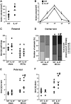
Increased chronic lesion volumes and worse functional outcome in IL-6−/− mice after mild cerebral ischaemia. Neuronal damage was assessed at 4 weeks after MCAO/reperfusion using NeuN immunohistochemistry. Cerebral lesion volumes are presented as individual data points (A). Direct cerebral lesion areas (B) were determined on five coronal brain sections (approximately interaural 6.6, 5.3, 3.9, 1.9 and -0.1 mm, respectively) by computer-assisted volumetry. Note that IL-6−/− mice show significantly enlarged chronic lesion volumes. *P < 0.05 versus wild-type. (C) Independent of genotype, MCAO animals showed worse Rotarod performance at 72 h post-stroke. (D) Behavioural asymmetries were assessed at 22 days after MCAO on the corner test. (E) Time to turn and (F) time to reach the floor at 23 days after MCAO on the pole test. Values are presented as individual data points (C, E and F) or as the percentage of animals that had performed the respective number of left turns (D). Horizontal bars = means. *P < 0.05 relative to sham, #P < 0.05 within genotypes. WT = wild-type.

References

    1. Acalovschi D, Wiest T, Hartmann M, Farahmi M, Mansmann U, Auffarth GU, et al. Multiple levels of regulation of the interleukin-6 system in stroke. Stroke. 2003;34:1864–9. - PubMed
    1. Andjelkovic AV, Stamatovic SM, Keep RF. The protective effects of preconditioning on cerebral endothelial cells in vitro. J Cereb Blood Flow Metab. 2003;23:1348–55. - PubMed
    1. Azari BM, Marmur JD, Salifu MO, Ehrlich YH, Kornecki E, Babinska A. Transcription and translation of human F11R gene are required for an initial step of atherogenesis induced by inflammatory cytokines. J Transl Med. 2011;9:98. - PMC - PubMed
    1. Bromberg J, Darnell JE., Jr The role of STATs in transcriptional control and their impact on cellular function. Oncogene. 2000;19:2468–73. - PubMed
    1. Carmeliet P, Lampugnani MG, Moons L, Breviario F, Compernolle V, Bono F, et al. Targeted deficiency or cytosolic truncation of the VE-cadherin gene in mice impairs VEGF-mediated endothelial survival and angiogenesis. Cell. 1999;98:147–57. - PubMed

Publication types

MeSH terms