. 2012 Aug;40(14):6673-82.
doi: 10.1093/nar/gks314. Epub 2012 Apr 9.

Combinatorial targeting of ribbon-helix-helix artificial transcription factors to chimeric recognition sites

Affiliations

Combinatorial targeting of ribbon-helix-helix artificial transcription factors to chimeric recognition sites

Massimiliano Zampini et al. Nucleic Acids Res. 2012 Aug.

Abstract

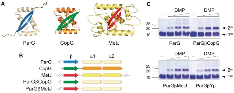
Artificial transcription factors (ATFs) are potent synthetic biology tools for modulating endogenous gene expression and precision genome editing. The ribbon-helix-helix (RHH) superfamily of transcription factors are widespread in bacteria and archaea. The principal DNA binding determinant in this family comprises a two-stranded antiparallel β-sheet (ribbons) in which a pair of eight-residue motifs insert into the major groove. Here, we demonstrate that ribbons of divergent RHH proteins are compact and portable elements that can be grafted into a common α-helical scaffold producing active ATFs. Hybrid proteins cooperatively recognize DNA sites possessing core tetramer boxes whose functional spacing is dictated by interactions between the α-helical backbones. These interactions also promote combinatorial binding of chimeras with different transplanted ribbons, but identical backbones, to synthetic sites bearing cognate boxes for each protein either in vitro or in vivo. The composite assembly of interacting hybrid proteins offers potential advantages associated with combinatorial approaches to DNA recognition compared with ATFs that involve binding of a single protein. Moreover, the new class of RHH ATFs may be utilized to re-engineer transcriptional circuits, or may be enhanced with affinity tags, fluorescent moieties or other elements for targeted genome marking and manipulation in bacteria and archaea.


Figures

Figure 1.
Figure 1.
Organization of chimeric ParG:RHH proteins. (A) Tertiary structures of ParG (1P94.pdb), MetJ (1CMA.pdb) and CopG (2CPG.pdb) dimers. α-helices are shown in shades of orange and β-strands in blue (ParG), green (CopG) and red (MetJ). (B) Linear representations of ParG, CopG, MetJ and chimeras ParGβCopG and ParGβMetJ that comprise ribbons of CopG and MetJ, respectively, transplanted into the ParG α-helical scaffold. (C) Chemical cross-linking of ParG, ParGβCopG, ParGβMetJ and ParGβYp with dimethyl pimelimidate (0, 1, 5 and 10 mM, left to right) at 37°C for 60 min followed by SDS–PAGE. Monomeric (1m) and dimeric (2m) species, and positions of molecular weight markers (kDa) are shown.
Figure 2.
Figure 2.
Docking of ParGβCopG and ParGβMetJ onto hybrid sites. (A) The popG locus comprises a site for ParG binding in which 5′-ACTC-3′ boxes were replaced by 5′-GCAC-3′ core motifs recognized by ParGβCopG. The hybrid was tested in gel retardation assays with double-stranded oligonucleotides bearing popG, and with variant sites in which T residues were introduced 5′ of the tetramer motifs (popG1), left and right 5′-GCAC-3′ tetramers were inverted (popG2 and popG3, respectively), and AT contents of regions flanking the 5′-GCAC-3′ boxes were decreased (popG4). Open and filled arrows mark unbound DNA and nucleoprotein complexes, respectively. (B) Sequences derived from sites recognized by ParG and MetJ are represented by blue and red boxes, respectively. The pet1 site bears the sequence recognized by MetJ in the crystal structure; pet2 is similar to pet1 but carries a C-to-T mutation in the spacer between the two 5′-ACGT-3′ boxes; pet3 consists of a ParG site in which core 5′-ACTC-3′ motifs recognized by the protein are replaced with 5′-ACGT-3′ sequences for ParGβMetJ binding. The control is a 24-bp double-stranded oligonucleotide with a randomized sequence. Open and filled arrows mark unbound DNA and nucleoprotein complexes, respectively. (C) Testing ribbon sequence requirements for ParGβCopG binding. The ParG ribbon comprises KRVNVNFD that is altered to KRLTITLS in ParGβCopG which possesses the CopG ribbon (green). Amino acids one and two are common to parental and hybrid proteins. In ParGβCopG.1, residues 3–6 correspond to those in CopG, whereas residues 4 and 6 in ParGβCopG.2 are derived from CopG. Other residues originate from ParG. ParGβCopG.1 and ParGβCopG.2 were tested in gel retardation assays with the popG site. Proteins in panels A–C were used at 0, 0.5, 1.0 and 1.5 µM (left to right).
Figure 3.
Figure 3.
Design and binding of hybrids with ribbon motifs from putative RHH proteins. (A) Left: Sequences of the ParG β-strand and putative ribbons from homologues in Y. pseudotuberculosis and A. salmonicida (GenBank accession numbers CAQ76574 and CAQ81938, respectively). Differences from ParG are highlighted in magenta and black. These motifs were swapped into ParG to generate hybrids ParGβYp and ParGβAs. Right: Organization of 5′-ACTC-3′ boxes in the OF site bound by ParG (15), and 5′-GAC/TA-3′ and 5′-CATT/G-3′ motifs in the Y- and A-sites of Y. pseudotuberculosis and A. salmonicida, respectively. Arrow orientation indicates direct or inverted boxes. (B) Gel retardation assays with OF, the Y-site, ParG and ParGYp. (C) Gel retardation assays with OF, the Y-site, and ParGβYp. Assays in panels (B) and (C) used 0, 0.3, 0.4, 0.5, 0.6, 0.7, 0.8, 1.0 and 1.5 µM (left to right) of the indicated proteins. (D) Gel retardation assays with OF, the A-site, and ParGβAs. Assays used 0, 0.1, 0.2, 0.3, 0.4 and 0.5 µM (left to right) ParGβAs. Open and filled arrows mark unbound DNA and nucleoprotein complexes, respectively, in panels A–D.
Figure 4.
Figure 4.
Simultaneous binding of ParG and hybrid ParGβCopG to chimeric sites. (A) Sequences of binding sites for ParG, CopG and hybrid sites tested for binding by ParG and/or ParGβCopG. Sequences from sites recognized by ParG and CopG are highlighted in blue and green, respectively. Core recognition motifs for ParG and ParGβCopG—an inverted 5′-ACTC-3′ sequence and 5′-GCAC/T-3′, respectively—are boxed. (B) Gel retardation assays used proteins mixed in equimolar ratios to give final concentrations of 0, 0.5, 1.0 and 1.5 µM (left to right) that were analyzed for binding to the copGLparGR and parGLcopGR sites (left panels). In addition, sites harboring single base-pair deletions or insertions in the central AT-rich regions were tested (middle and right panels, respectively). Open and filled arrows mark unbound DNA and nucleoprotein complexes, respectively. (C) Surface plasmon resonance analysis of ParG and ParGβCopG binding individually and simultaneously to the copGLparGR site. (D) DNase I footprinting of ParG and ParGβCopG on copGLparGR. Proteins were mixed in equimolar ratios to give final concentrations of 0, 3, 9, 16 and 20 µM (left to right). The location of the copGLparGR site is highlighted. L, Maxam–Gilbert A + G sequencing ladder.
Figure 5.
Figure 5.
Chimera ParGβCopG is a functional transcriptional repressor in vivo. (A) The regulatory region (POF) for parF-parG expression comprises a characteristic Gram-negative promoter in which an array of eight 5′-ACTC-3′ boxes (blue) recognized by ParG is situated downstream of the putative −10 motif (15). These tetramer boxes were replaced by 5′-GCAC-3′ motifs (green) derived from a copG site (Figure 1) in the synthetic POH1 promoter-operator, and by alternating 5′-GCAC-3′ and 5′-ACGT-3′ boxes (red) in POH2—the latter are recognized by ParGβMetJ. (B) Synthetic promoter-operators were cloned as transcriptional fusions to a lacZ gene in pRS415. Intrinsic promoter activities when empty pCDFDuet-1 was present in trans and repression of these activities by ParGβCopG and/or ParGβMetJ produced from pCDFDuet-1 were tested. (C) Resensitization of E. coli to kanamycin. Native regulatory signals controlling expression of aph were supplanted by the POH1 promoter-operator. ParGβCopG provided in trans from a compatible pCDFDuet-1 based vector repressed expression from POH1 and ablated kanamycin resistance (filled diamonds). ParGβMetJ alone (filled squares) and ParGβCopG and ParGβMetJ simultaneously (open triangles) repressed aph expression to intermediate levels. Open circles show growth of a strain bearing the POH1-aph fusion and empty pCDFDuet-1. The inset shows that strains carrying the POH1-aph fusion and either pCDFDuet-1 (open circles) or pCDFDuet-1 expressing ParGβCopG (filled diamonds) grew similarly. (D) Interaction between positively charged lysine at β-strand position one and negatively charged glutamate approaching from a nearby α-helix may assist in stabilizing the ribbon in ParG and functional hybrids. A similar interaction will occur at the other ribbon extremity.

Similar articles

Cited by

References

    1. Stormo GD, Zhao Y. Determining the specificity of protein-DNA interactions. Nat. Rev. Genet. 2010;11:751–760. - PubMed
    1. Garvie CW, Wolberger C. Recognition of specific DNA sequences. Mol. Cell. 2001;8:937–946. - PubMed
    1. Ansari AZ, Mapp AK. Modular design of artificial transcription factors. Curr. Opin. Chem. Biol. 2002;6:765–772. - PubMed
    1. Collins CH, Yokobayashi Y, Umeno D, Arnold FH. Engineering proteins that bind, move, make and break DNA. Curr. Opin. Biotechnol. 2003;14:371–378. - PubMed
    1. Khalil AS, Collins JJ. Synthetic biology: applications come of age. Nat. Rev. Genet. 2010;11:367–379. - PMC - PubMed

Publication types