. 2012 Jul;97(7):E1316-26.
doi: 10.1210/jc.2012-1018. Epub 2012 Apr 6.

Endometrial miR-181a and miR-98 expression is altered during transition from normal into cancerous state and target PGR, PGRMC1, CYP19A1, DDX3X, and TIMP3

Affiliations

Endometrial miR-181a and miR-98 expression is altered during transition from normal into cancerous state and target PGR, PGRMC1, CYP19A1, DDX3X, and TIMP3

Harekrushna Panda et al. J Clin Endocrinol Metab. 2012 Jul.

Abstract

Context: Evidence suggests that a number of microRNA (miRNA) are aberrantly expressed in endometrial disorders with potential posttranscriptional regulation of their specific target genes, including ovarian steroid receptors.

Objectives: Our objective was to assess the endometrial expression of miR-98 and miR-181a and their respective target genes, progesterone (P4) receptor membrane component 1 (PGRMC1) and P4 receptor (PGR).

Design, setting, and patients: We evaluated tissue expression and in vitro regulation at an academic university medical center in endometrial biopsies and endometrial tissues from follicular and luteal phases with and without exposure to hormonal therapies and grade I-III endometrial cancer (n = 52).

Interventions: INTERVENTIONS included endometrial biopsies and in vitro transfection.

Main outcome measures: We evaluated expression and function of miR-98 and miR-181a.

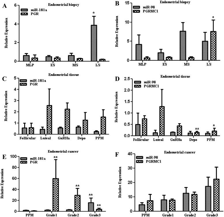
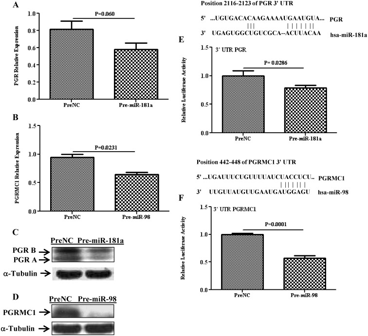
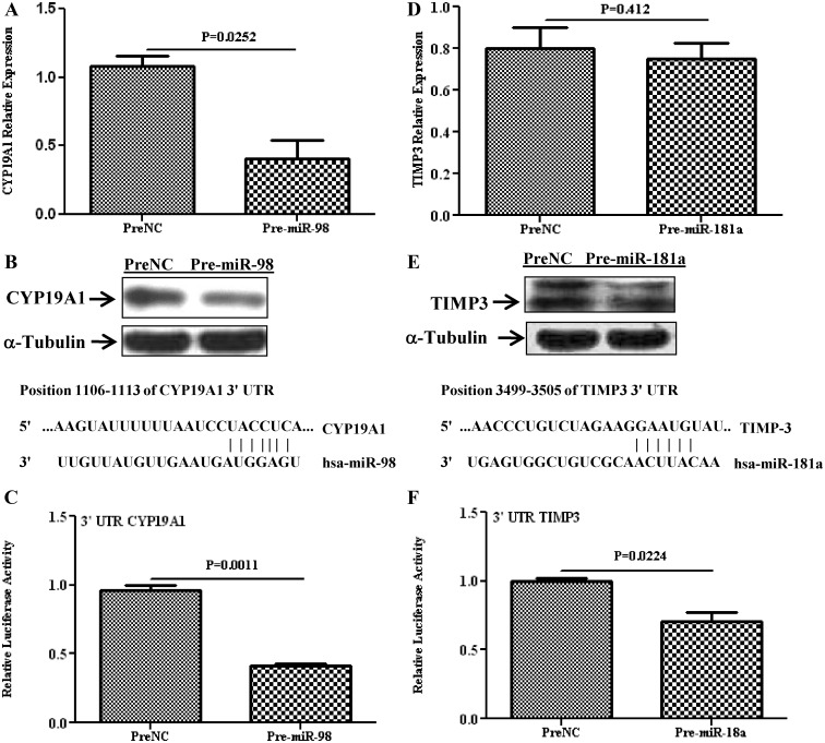
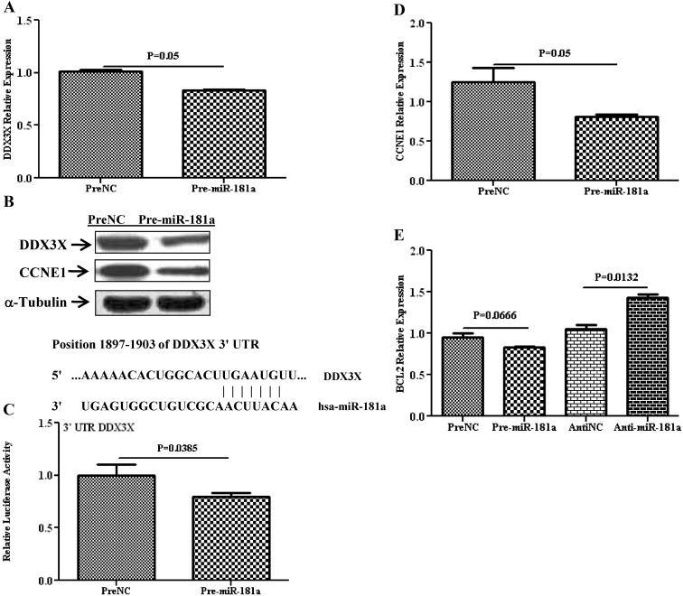
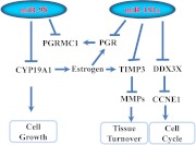
Results: Aberrant expression of miR-98 and miR-181a is associated with endometrial transition from normal into cancerous states, which to some extent is influenced by hormonal milieu, and exhibited an inverse relationship with PGMRC1 and PGR expression, respectively. Treatments of Ishikawa cells with 17β-estradiol, P4, or medroxyprogesterone acetate had limited effects on miR-98, miR-181a, and PGRMC1 expression, whereas 17β-estradiol treatment increased PGR expression. In Ishikawa cells, gain of function of miR-98 repressed PGRMC1 and CYP19A1, and miR-181a repressed PGR, DDX3X, and TIMP3 at mRNA and protein levels through direct interactions with their respective 3'-untranslated regions and CCNE1 through miR-181a-induced DDX3X repression, with miR-98 reducing the rate of cell proliferation as compared with controls.

Conclusion: miR-98 and miR-181a through their regulatory functions on PGRMC1, PGR, CYP19A1, TIMP3, and DDX3X expression may influence a wide range of endometrial cellular activities during normal menstrual cycle and transition into disease states, including endometrial cancer.


Figures

Fig. 1.
Fig. 1.
Relative expression of miR-98, miR-181a, PGR, and PGRMC1 in endometrial biopsies (A and B) from mid-late proliferative (MLP), early-secretory (ES), midsecretory (MS), and late-secretory (LS) phases of the menstrual cycle and endometrial tissues (C and D) from follicular and luteal phases of the menstrual cycle and peri-postmenopausal period (PPM) from women who were exposed to GnRHa and Depo-Provera (Depo) as well as endometrial cancer (E and F) grades I, II, and III. During the calculation of expression value, one of the samples from MLP, follicular phase, and PPM was taken as control for endometrial biopsies, endometrial tissues, and endometrial cancer, respectively. The values are reported as mean ± sem and analyzed using nonparametric t test and ANOVA. *, P < 0.05; **, P < 0.005, indicating significant difference from the control.
Fig. 2.
Fig. 2.
The effect of E2, P4, and MPA on the expression of miR-98, miR-181a, PGR, and PGRMC1 in Ishikawa cells compared with untreated control [control (Ctrl)], after 6, 24, and 48 h of treatments as determined by quantitative RT-PCR. The results are presented as mean ± sem of three independent experiments and analyzed using nonparametric Student's t test. *, P < 0.05, indicating significantly different from untreated control.
Fig. 3.
Fig. 3.
Relative expression of PGR and PGRMC1 in Ishikawa cells after overexpression of miR-181a and miR-98 (pre-miR-transfected) as compared with cells transfected with PreNC determined by quantitative RT-PCR (A and B) and Western blot analysis (C and D). Firefly luciferase assay with pGL3-TK modified construct and pZEX-MT01 construct carrying a 3′-UTR fragment of PGR (E) and PGRMC1 (F), respectively, in which Ishikawa cells were cotransfected with, respectively, firefly luciferase construct, pre-miR-181a, or pre-miR-98 or PreNC. The ratio of firefly to renilla was determined and reported as relative luciferase activity and was compared with levels in PreNC, which was set at 1. The results present as mean ± sem of three sets of independent experiments performed in duplicate and analyzed using nonparametric Student's t test with P values presented as indicated by corresponding line. Sequence alignment of miR-181a and miR-98 seed regions and PGR and PGRMC1 mRNA target site at their 3′-UTR with the coordinated positions are shown at the top of each graph.
Fig. 4.
Fig. 4.
Relative expression of CYP19A1 and TIMP3 in Ishikawa cells after overexpression of miR-98 and miR-181a (pre-miR-transfected), respectively, as compared with PreNC determined by quantitative RT-PCR (A and D) and Western blot analysis (B and E). Firefly luciferase assay with pZEX-MT01 construct and pIS0 construct carrying a 3′-UTR fragment of CYP19A1 (C) and TIMP3 (F), respectively, in Ishikawa cells cotransfected with firefly luciferase construct, pre-miR-98 or pre-miR-181a or preNC. The ratio of firefly to renilla was determined and reported as relative luciferase activity and was compared with levels in PreNC, which was set at 1. The results present mean ± sem of three sets of independent experiments performed in duplicate and analyzed using nonparametric Student's t test with P values presented as indicated by corresponding line. Sequence alignment of miR-181a and miR-98 seed regions and CYP19A1 and TIMP3 mRNA target site at their 3′-UTR with the coordinated positions are shown at the top of each graph.
Fig. 5.
Fig. 5.
Relative expression of DDX3X and CCNE1 in Ishikawa cells after overexpression of miR-181a (pre-miR-181a), or PreNC determined by quantitative RT-PCR (A and D) and Western blot analysis (B). C, Firefly luciferase assay with pZEX-MT01 construct carrying a 3′-UTR fragments of DDX3X in Ishikawa cells cotransfected with firefly luciferase reporter, pre-miR-181a, or PreNC. The ratio of firefly to renilla was determined and reported as relative luciferase activity compared with empty vector with levels in PreNC independently set at 1. The results present mean ± sem of three sets of independent experiments performed in duplicate and analyzed using nonparametric Student's t test with P values presented as indicated by corresponding line. Sequence alignment of miR-181a seed region and DDX3X mRNA target site at 3′-UTR with the coordinated positions is shown at the top of each graph. E, Relative expression of BCL2 in Ishikawa cells after overexpression of miR-181a (Pre-miR-181a) as compared to its control (PreNC) and depletion of miR-181a (Anti-miR-181a) as compared to its control (AntiNC) by quantitative RT-PCR.
Fig. 6.
Fig. 6.
Schematic presentation of miR-98 and miR-181a regulatory function on specific target genes, including PGR, PGRMC1, CYP19A1, TIMP3, and DDX3X and the regulatory interactions among their products in endometrial cells. The expression and regulatory interactions among these gene products regulated by miR-98 and miR-181a may influence various cellular activities that result in tissue turnover, cell cycle progression, and growth.

Similar articles

Cited by

References

    1. Bartel DP. 2009. MicroRNA: target recognition and regulatory functions. Cell 136:215–233 - PMC - PubMed
    1. Djuranovic S, Nahvi A, Green R. 2011. A parsimonious model for gene regulation by miRNA. Science 331:550–553 - PMC - PubMed
    1. Fabian MR, Sonenberg N, Filipowicz W. 2010. Regulation of mRNA translation and stability by microRNA. Annu Rev Biochem 79:351–379 - PubMed
    1. Almeida MI, Reis RM, Calin GA. 2011. MicroRNA history: Discovery, recent applications, and next frontiers. Mutat Res 717:1–8 - PubMed
    1. Hagen JW, Lai EC. 2008. microRNA control of cell-cell signaling during development and disease. Cell Cycle 7:2327–2332 - PMC - PubMed

Publication types

MeSH terms