. 2012 Jun;14(6):777-89.
doi: 10.1093/neuonc/nos077. Epub 2012 Apr 5.

Interplay among BRAF, p16, p53, and MIB1 in pediatric low-grade gliomas

Affiliations

Interplay among BRAF, p16, p53, and MIB1 in pediatric low-grade gliomas

Craig Horbinski et al. Neuro Oncol. 2012 Jun.

Abstract

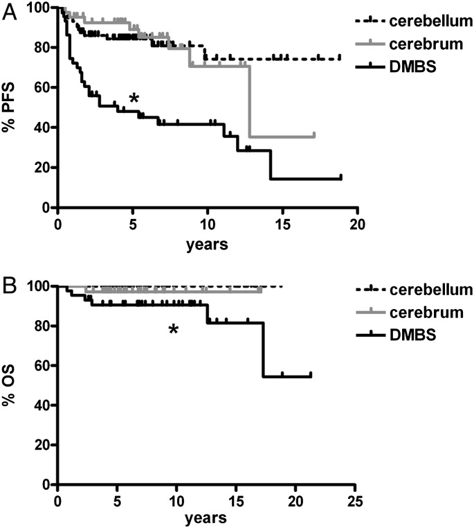
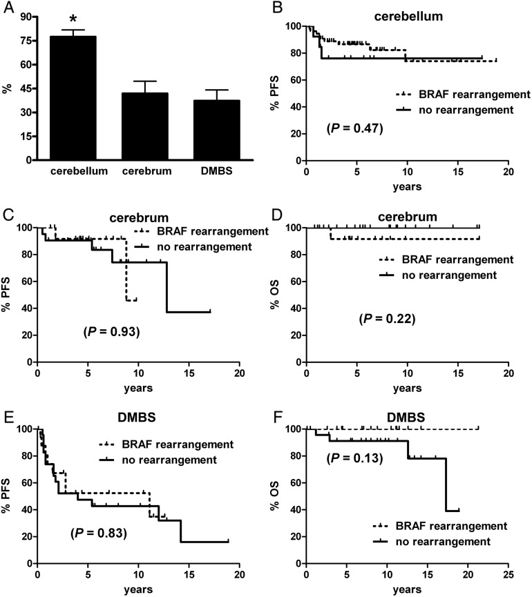
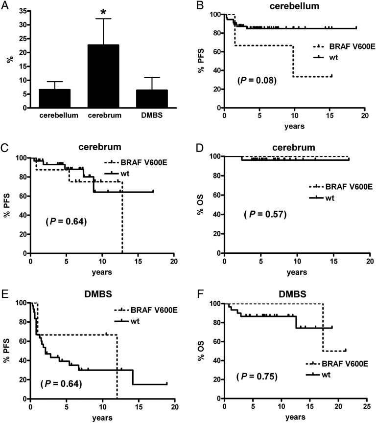
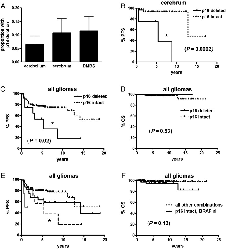
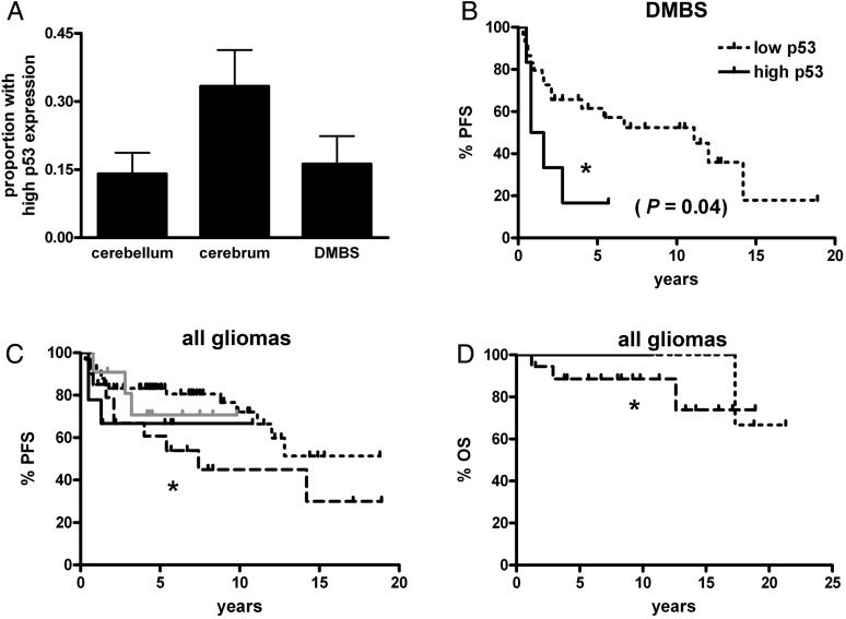
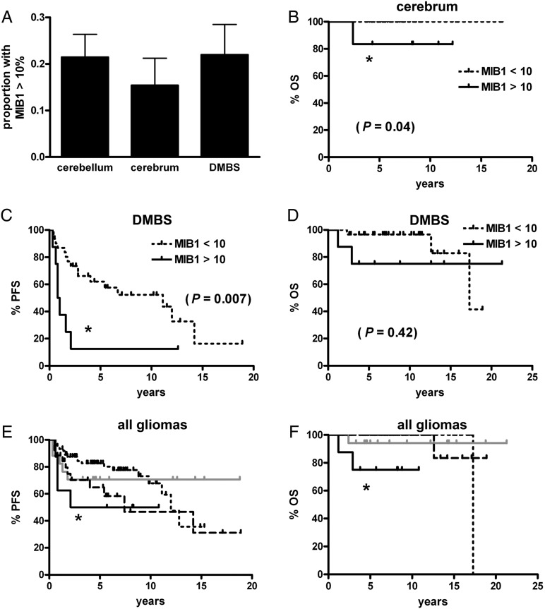
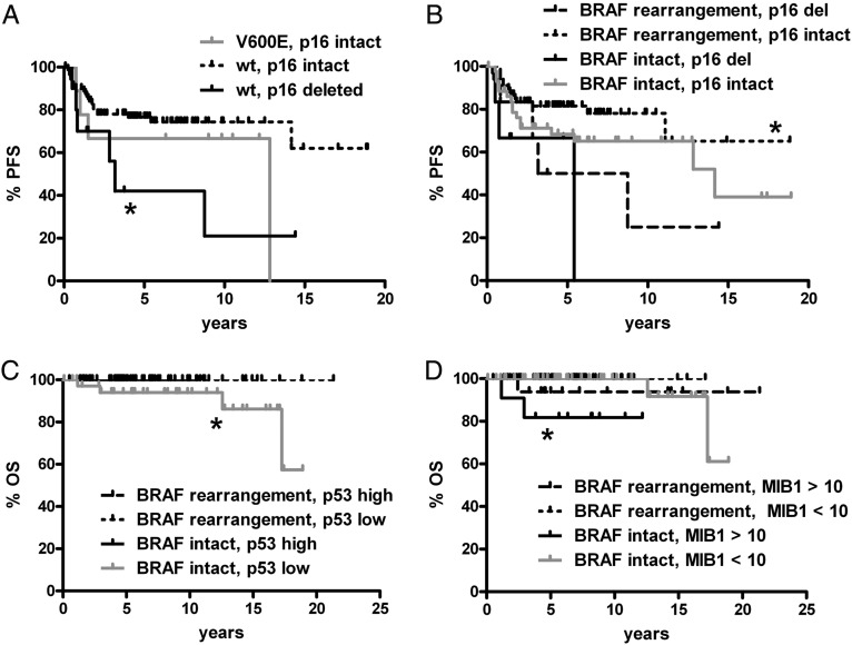
BRAF rearrangements and BRAF V600E point mutations are recurring events in pediatric low-grade gliomas. However, their clinical significance, including possible interactions between these markers and other glioma biomarkers, is unclear. In this study a retrospective cohort of 198 pediatric low-grade gliomas (including 40 treated with adjuvant therapy) was analyzed for BRAF rearrangements, BRAF V600E, p16/CDKN2A deletion, p53 expression, and MIB1 proliferation index. In tumors with BRAF rearrangement, homozygous p16 deletion correlated with shorter progression-free survival (P = .04). A high MIB1 proliferation index trended toward worse response to adjuvant radiotherapy compared to BRAF-rearranged, p16-intact tumors (P = .08). On multivariate analysis, the 2 most consistently powerful independent adverse prognostic markers were midline location (P = .0001) and p16 deletion (P = .03). Tumors with BRAF V600E had a strong trend toward an increased risk for progression (hazard ratio = 2.48, P = .07), whereas those with BRAF rearrangement had a milder trend toward reduced risk (hazard ratio = .54, P = .15). These data suggest that p16 deletion adversely impacts the outcomes of BRAF-driven gliomas, that high proliferation index may be a better marker of progression risk than BRAF, that BRAF rearrangement and BRAF V600E might not necessarily produce comparable outcomes, and that none of these markers is stronger than tumor location in determining prognosis in pediatric low-grade gliomas.


Figures

Fig. 1.
Fig. 1.
Progression-free and overall survival in pediatric low-grade gliomas are worse in midline locations. Tumors arising in the midline (ie, DMBS) had generally worse progression-free survival (A) and overall survival (B) compared with those located in the cerebellum and cerebrum. In (A), *P < .0001 vs cerebellum and = .0003 vs cerebrum; in (B) *P < .001 vs cerebellum and = .17 vs cerebrum.
Fig. 2.
Fig. 2.
PFS and OS in pediatric low-grade gliomas by location and BRAF rearrangement status. (A) Tumors in the cerebellum had a higher rate of BRAF rearrangement than did those in the cerebrum or midline, aka DMBS. *P < .001 via Kruskal–Wallis and Dunn's post hoc. (B–F) BRAF rearrangement did not significantly stratify PFS or OS in any of the 3 tumor subgroups. No deaths occurred in any cerebellar tumor.
Fig. 3.
Fig. 3.
PFS and OS in pediatric low-grade gliomas by location and BRAF V600E status. (A) Tumors in the cerebrum had a higher rate of BRAF V600E mutation than those in the cerebellum (*P < .05) or midline, aka DMBS. While there was a trend toward worse PFS in cerebellar tumors with BRAF V600E (B), BRAF V600E did not significantly stratify PFS or OS in any of the other tumor subgroups (C–F). No deaths occurred in any cerebellar tumor.
Fig. 4.
Fig. 4.
PFS and OS in pediatric low-grade gliomas by p16 and BRAF status. (A) There was no difference in the frequency of homozygous p16 deletion by tumor site (P = .64). While PFS was shorter in cerebral tumors (B) and all pooled gliomas (C) with p16 deletion, cerebellar and DMBS subsets did not achieve significance (see Results), nor did OS in all gliomas (D). (E) Stratifying all gliomas by p16 deletion and any BRAF abnormality (rearrangement or V600E mutation) showed that when BRAF was abnormal, the concomitant presence of p16 deletion correlated with shorter PFS (short dotted black line = p16 intact, BRAF abnormal; long dotted black line = p16 deleted, BRAF abnormal; solid grey line = p16 deleted, BRAF normal; solid black line = p16 intact, BRAF normal; *P = .04 vs p16 intact, BRAF abnormal). (F) There was, however, no significant stratification of OS by p16 and BRAF in this cohort. DMBS, diencephalon/midbrain/brainstem/spinal cord; nl, normal.
Fig. 5.
Fig. 5.
PFS and OS in pediatric low-grade gliomas by p53 and BRAF status. (A) There was a strong trend toward the cerebrum having more high-expressing p53 tumors than either cerebellar or DMBS subregions (P = .06), although it was only in the DMBS subregion that high-expressing p53 tumors had shorter PFS (B). (C and D) In all pooled gliomas, those with weak p53 expression and normal BRAF had shorter PFS (C, *P = .05) and OS (D, *P = .02) than weak p53-expressing tumors with any BRAF abnormality (either rearrangement or V600E mutation). However, the degree of p53 expression did not modify PFS or OS when adjusting for BRAF status. Short dotted black line = p53 weak, BRAF abnormal; long dotted black line = p53 weak, BRAF normal; solid grey line = p53 strong, BRAF abnormal; solid black line = p53 strong, BRAF normal. DMBS, diencephalon/midbrain/brainstem/spinal cord.
Fig. 6.
Fig. 6.
PFS and OS in pediatric low-grade gliomas by MIB1 proliferation index and BRAF status. (A) There was no difference in the frequency of high proliferation index by tumor site (P = .70). OS was shorter in cerebral tumors with elevated MIB1 (B), as was PFS (but not OS) in DMBS tumors (C and D, respectively). No other comparisons within each subregion were significant. (E) Stratifying all gliomas by MIB1 and any BRAF abnormality (rearrangement or V600E mutation) showed that when MIB1 was high and BRAF was normal, PFS was shorter compared with tumors with low MIB1 and abnormal BRAF (*P = .049). (F) Likewise, gliomas with high MIB1 and normal BRAF had shorter OS compared with tumors with low MIB1 and abnormal BRAF (*P < .0001), as well as compared with tumors with low MIB1 and normal BRAF (*P = .02). Short dotted black line = MIB1 < 10%, BRAF abnormal; long dotted black line = MIB1 < 10%, BRAF normal; solid grey line = MIB1 >10%, BRAF abnormal; solid black line = MIB1 >10%, BRAF normal. DMBS, diencephalon/midbrain/brainstem/spinal cord.
Fig. 7.
Fig. 7.
Significant PFS and OS associations in pediatric low-grade gliomas by BRAF V600E or rearrangement and p16, p53, or MIB1. All subregions were pooled together; all comparisons not marked with an asterisk (or not shown) are not significant. (A) Tumors without BRAF V600E (wild type [wt] BRAF) but with p16 deletion had shorter PFS than wt BRAF tumors where p16 was intact (*P = .02 vs V600E, p16 intact). There was only 1 case with concomitant V600E mutation and p16 deletion—a left temporal PXA that progressed after 5.4 years, though the patient is alive 10.3 years after original surgery. (B) Likewise, tumors that had both BRAF rearrangement and intact p16 had better PFS than those with p16 deletion regardless of BRAF status (*P = .049 vs BRAF rearrangement, p16 deleted; *0.03 vs BRAF intact, p16 deleted). (C) OS (*P = .03) was shorter in gliomas with intact BRAF and low p53 expression compared with those with low p53 and BRAF rearrangement. PFS also showed a strong trend in the same direction (P = .06, not shown). (D) Tumors with intact BRAF and high MIB1 proliferation index had shorter OS than tumors with a lower proliferation index and either intact or rearranged BRAF (*P = .007 and .0008, respectively). There was also a trend toward worse OS in BRAF-rearranged tumors when MIB1 was high compared with BRAF-rearranged tumors with low MIB1 (P = .06). Del, deleted.

References

    1. Bar EE, Lin A, Tihan T, Burger PC, Eberhart CG. Frequent gains at chromosome 7q34 involving BRAF in pilocytic astrocytoma. J Neuropathol Exp Neurol. 2008;67:878–887. doi:10.1097/NEN.0b013e3181845622. - DOI - PubMed
    1. Pfister S. BRAF gene duplication constitutes a mechanism of MAPK pathway activation in low-grade astrocytomas. J Clin Invest. 2008;118:1739–1749. doi:10.1172/JCI33656. - DOI - PMC - PubMed
    1. Jones DT, Kocialkowski S, Liu L, et al. Tandem duplication producing a novel oncogenic BRAF fusion gene defines the majority of pilocytic astrocytomas. Cancer Res. 2008;68(21):8673–8677. doi:10.1158/0008-5472.CAN-08-2097. - DOI - PMC - PubMed
    1. Forshew T, Tatevossian RG, Lawson AR, et al. Activation of the ERK/MAPK pathway: a signature genetic defect in posterior fossa pilocytic astrocytomas. J Pathol. 2009;218(2):172–181. doi:10.1002/path.2558. - DOI - PubMed
    1. Jones DT, Kocialkowski S, Liu L, Pearson DM, Ichimura K, Collins VP. Oncogenic RAF1 rearrangement and a novel BRAF mutation as alternatives to KIAA1549:BRAF fusion in activating the MAPK pathway in pilocytic astrocytoma. Oncogene. 2009;28(20):2119–2123. - PMC - PubMed

Publication types

MeSH terms