

Use of magnetic perfusion-weighted imaging to determine epidermal growth factor receptor variant III expression in glioblastoma
- PMID: 22492960
- PMCID: PMC3337316
- DOI: 10.1093/neuonc/nos073
Use of magnetic perfusion-weighted imaging to determine epidermal growth factor receptor variant III expression in glioblastoma
Abstract
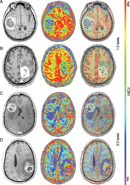
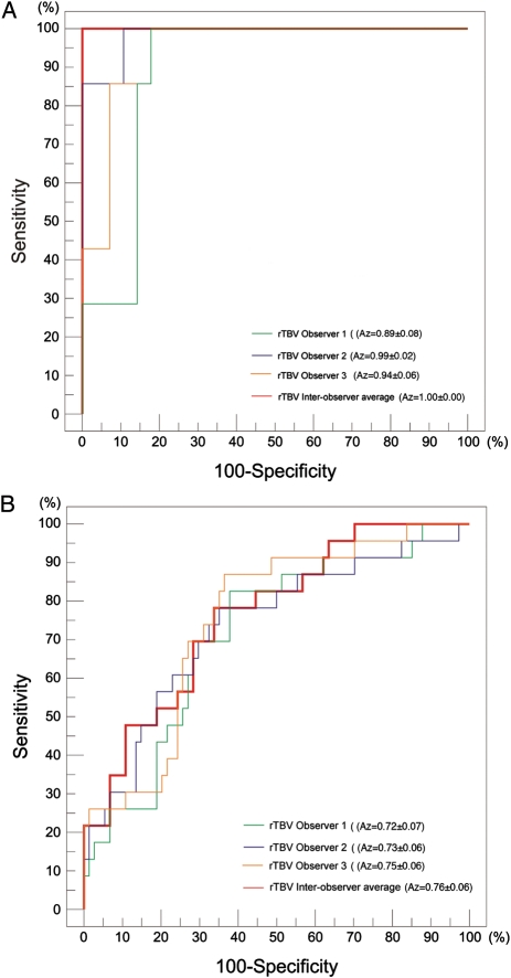
Identification of the epidermal growth factor receptor variant III (EGFRvIII) mutation in glioblastoma has become increasingly relevant in the optimization of therapy. Traditionally, determination of tumor EGFRvIII-expression has relied on tissue-based diagnostics. Here, we assess the accuracy of magnetic resonance perfusion-weighted imaging (MR-PWI) in discriminating the EGFRvIII-expressing glioblastoma subtype. We analyzed RNA from 132 primary human glioblastoma tissue samples by reverse-transcription polymerase chain reaction (RT-PCR) for the EGFRvIII and EGFR wild-type mutations and by quantitative RT-PCR for expression of vascular endothelial growth factor (VEGF). Concurrently, 3 independent observers reviewed preoperative 1.5-Tesla (T)/SE or 3.0-Tesla (T)/GE MR perfusion images to determine the maximum relative tumor blood volume (rTBV) of each of these tumors. EGFRvIII-expressing glioblastomas showed significantly higher rTBV, compared with those tumors lacking EGFRvIII expression. This association was observed in both the 1.5T/SE (P = .000) and 3.0T/GE (P = .001) cohorts. By logistic regression analysis, combining the 2 MR system cohorts, rTBV was a very strong predictor of EGFRvIII mutation (odds ratio [rTBV] = 2.70; P = .000; McFadden's ρ(2) = 0.23). Furthermore, by receiver-operating characteristic curve analysis, rTBV discriminated EGFRvIII with very high accuracy (A(z) = 0.81). In addition, we found that VEGF upregulation was associated, although without reaching statistical significance, with EGFRvIII expression (P = .16) and with increased rTBV (F-ratio = 2.71; P = .102). These trends suggest that VEGF-mediated angiogenesis may be a potential mediator of angiogenesis to increase perfusion in EGFRvIII-expressing glioblastomas, but there are likely several other contributing factors. This study demonstrates the potential to use rTBV, a MR-PWI-derived parameter, as a noninvasive surrogate of the EGFRvIII mutation.
Figures
References
-
- Krex D, Klink B, Hartmann C, et al. Long-term survival with glioblastoma multiforme. Brain. 2007;130(pt 10):2596–2606. doi:10.1093/brain/awm204. - DOI - PubMed
-
- Bernays RL, Kollias SS, Khan N, Brandner S, Meier S, Yonekawa Y. Histological yield, complications, and technological considerations in 114 consecutive frameless stereotactic biopsy procedures aided by open intraoperative magnetic resonance imaging. J Neurosurg. 2002;97(2):354–362. doi:10.3171/jns.2002.97.2.0354. - DOI - PubMed
-
- Arvinda HR, Kesavadas C, Sarma PS, et al. Glioma grading: sensitivity, specificity, positive and negative predictive values of diffusion and perfusion imaging. J Neurooncol. 2009;94(1):87–96. doi:10.1007/s11060-009-9807-6. - DOI - PubMed
-
- Gasparetto EL, Pawlak MA, Patel SH, et al. Posttreatment recurrence of malignant brain neoplasm: accuracy of relative cerebral blood volume fraction in discriminating low from high malignant histologic volume fraction. Radiology. 2009;250(3):887–896. doi:10.1148/radiol.2502071444. - DOI - PubMed
Publication types
MeSH terms
Substances
Grants and funding
LinkOut - more resources
Full Text Sources
Other Literature Sources
Medical
Research Materials
Miscellaneous
