. 2012 Apr 24;109(17):6584-9.
doi: 10.1073/pnas.1113271109. Epub 2012 Apr 5.

ErbB receptor tyrosine kinase/NF-κB signaling controls mammosphere formation in human breast cancer

Affiliations

ErbB receptor tyrosine kinase/NF-κB signaling controls mammosphere formation in human breast cancer

Kunihiko Hinohara et al. Proc Natl Acad Sci U S A. .

Abstract

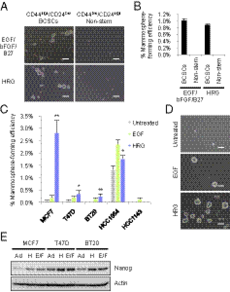
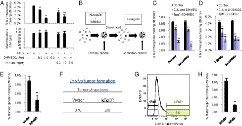
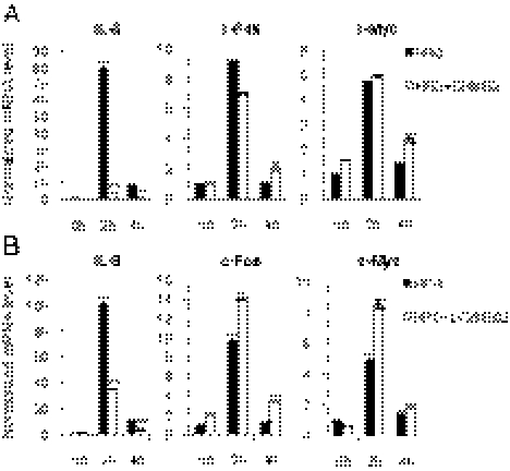
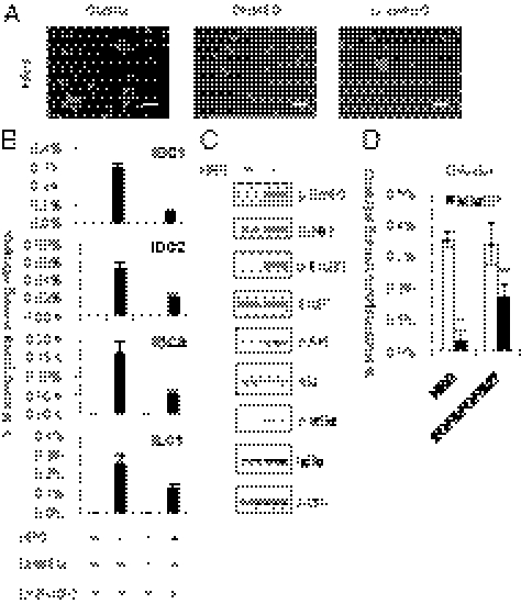
Breast cancer is one of the most common cancers in humans. However, our understanding of the cellular and molecular mechanisms underlying tumorigenesis in breast tissues is limited. Here, we identified a molecular mechanism that controls the ability of breast cancer cells to form multicellular spheroids (mammospheres). We found that heregulin (HRG), a ligand for ErbB3, induced mammosphere formation of a breast cancer stem cell (BCSC)-enriched population as well as in breast cancer cell lines. HRG-induced mammosphere formation was reduced by treatment with inhibitors for phosphatidyl inositol 3-kinase (PI3K) or NF-κB and by expression of IκBα-Super Repressor (IκBαSR), a dominant-negative inhibitor for NF-κB. Moreover, the overexpression of IκBαSR in breast cancer cells inhibited tumorigenesis in NOD/SCID mice. Furthermore, we found that the expression of IL8, a regulator of self-renewal in BCSC-enriched populations, was induced by HRG through the activation of the PI3K/NF-κB pathway. These findings illustrate that HRG/ErbB3 signaling appears to maintain mammosphere formation through a PI3K/NF-κB pathway in human breast cancer.


Conflict of interest statement

The authors declare no conflict of interest.

Figures

Fig. 1.
Fig. 1.
Effect of HRG on mammosphere formation of a BCSC-enriched population. (A) Representative images of primary cultures of mammospheres formed from the sorted CD44high/CD24low/lineage BCSC-enriched population (Left) and the CD44low/CD24high/lineage nonstem cell population (Right) obtained from a specimen of invasive ductal carcinoma (IDC1, Table S1). The cells from IDC1 were incubated with EGF/bFGF/B27 (Top) or with 20 ng/mL HRG (Bottom). Scale bar = 100 μm. (B) The spheres were counted and the percentage of mammosphere-forming cells were determined in each group (data are mean ± SD; n = 4). (C) Mammosphere assay in MCF7, T47D, BT20, HCC1954, and HCC1143 breast cancer cell lines treated with 20 ng/mL EGF or 20 ng/mL HRG (data are mean ± SD; n = 4, **P < 0.01, *P < 0.05, relative to the values in the respective untreated controls). (D) Images showing mammosphere formation in MCF7 cells treated as indicated in (C). Scale bar = 100 μm. (E) Nanog protein expression levels in the parental cells growing in 2D adherent (Ad) culture, sphere cells cultured with 20 ng/mL HRG (H) and sphere cells cultured with EGF/bFGF/B27 (E/F).
Fig. 2.
Fig. 2.
HRG induces the ErbB/PI3K/Akt/NF-κB pathway. (A) MCF7, T47D, and BT20 cells were treated with 100 ng/mL HRG for 10 min, and protein expression levels were determined by immunoblotting. (B) MCF7 cells were treated with 100 ng/mL HRG for 10 min, and immunoprecipitation (IP) assays were performed with the indicated antibodies. (C) MCF7 cells were treated with 100 ng/mL HRG for 10, 30, and 60 min. Protein expression levels were determined by immunoblotting. (D) MCF7 cells were treated with 100 ng/mL HRG for 1, 2, 4, 8, and 16 h. The DNA binding activity of RELA was quantified by ELISA (data are mean ± SD; n = 3). (E) MCF7 cells were treated with 5 μg/mL DHMEQ (NF-κB inhibitor) or 10 μM LY294002 (PI3Kinase inhibitor) for 2 h, and then the cells were treated with 100 ng/mL HRG for 30, 60, and 90 min. The DNA-binding activity of RELA was quantified by ELISA (data are mean ± SD; n = 3).
Fig. 3.
Fig. 3.
Role of the HRG/PI3K/NF-κB axis on mammosphere formation. (A) MCF7 cells were incubated with 20 ng/mL HRG and/or the indicated concentrations of DHMEQ and LY294002. The number of formed mammospheres was counted, and the percentage of mammosphere-forming cells was determined [data are mean ± SD; n = 3, **P < 0.01, *P < 0.05, relative to the values in the HRG(+)]. (B) Experimental strategies for evaluating the effect of initial treatment with inhibitors on secondary mammosphere formation. (C and D) Effects of DHMEQ and LY294002 on primary and secondary mammosphere formation. MCF7 cells were incubated with 20 ng/mL HRG and/or the indicated concentrations of DHMEQ and LY294002. The formed primary mammospheres were dissociated into single cells and grown as secondary mammospheres without treatment with inhibitors. The mammospheres were counted, and the percentage of mammosphere-forming cells was determined (data are mean ± SD; n = 3, **P < 0.01, *P < 0.05, relative to the values in the respective controls). (E) MCF7 cells expressing the indicated lentiviral vectors were incubated with 20 ng/mL HRG, and the percentage of mammosphere-forming cells was determined (data are mean ± SD; n = 4, **P < 0.01). (F) NOD/SCID mice were injected in the mammary fat pad with 1 × 10⁵ of vector- or IκBαSR-transduced MCF7 cells. Tumor formation was indicated by tumors/injections at 6 wk after injection. (G) NF-κB reporter activity of mammospheres derived from MCF7 cells. (H) NF-κB+ cells and NF-κB cells (Fig. 3G) were sorted by FACS and cultured with 20 ng/mL HRG. The percentage of mammosphere-forming cells was determined (data are mean ± SD; n = 4, **P < 0.01).
Fig. 4.
Fig. 4.
IL8 is a transcriptional target of NF-κB. (A and B) MCF7 cells were treated with 5 μg/mL DHMEQ or 5 μM LY294002 for 2 hours, and then the cells were treated with 100 ng/mL HRG for 2 h and 4 h. Expression levels of IL8, c-Fos, and c-Myc were examined by quantitative RT-PCR (data are mean ± SD; n = 3, **P < 0.01, relative to the values in the respective controls).
Fig. 5.
Fig. 5.
The HRG/PI3K/NF-κB axis controls mammosphere formation of primary tumor cells derived from breast cancer patients. (A) Representative images of primary cultures of mammospheres incubated with 20 ng/mL HRG, 5 μg/mL DHMEQ and 5 μM LY294002. Scale bar = 100 μm. (B) Primary breast cancer cells obtained from specimens of invasive ductal carcinoma (IDC1, 2, and 3) or invasive lobular carcinoma (ILC1) were treated as indicated in A, and the percentage of mammosphere-forming cells was determined (data are mean ± SD; n = 4). (C) Cells from IDC1 were incubated with 100 ng/mL HRG for 10 min, and protein expression levels were determined by immunoblotting. (D) Cells from IDC1 expressing the indicated lentiviral vectors were incubated with 20 ng/mL HRG or with EGF/bFGF/B27, and the percentage of mammosphere-forming cells was determined (data are mean ± SD; n = 4, **P < 0.01, relative to the values in the respective controls).

References

    1. Reya T, Morrison SJ, Clarke MF, Weissman IL. Stem cells, cancer, and cancer stem cells. Nature. 2001;414:105–111. - PubMed
    1. Gotoh N. Potential signaling pathways activated in cancer stem cells in breast cancer. In: Shostak S, editor. Cancer Stem Cells Theories and Practice. Vienna, Austria: InTech; 2011. p. 6.
    1. Al-Hajj M, Wicha MS, Benito-Hernandez A, Morrison SJ, Clarke MF. Prospective identification of tumorigenic breast cancer cells. Proc Natl Acad Sci USA. 2003;100:3983–3988. - PMC - PubMed
    1. Boiko AD, et al. Human melanoma-initiating cells express neural crest nerve growth factor receptor CD271. Nature. 2010;466:133–137. - PMC - PubMed
    1. Bonnet D, Dick JE. Human acute myeloid leukemia is organized as a hierarchy that originates from a primitive hematopoietic cell. Nat Med. 1997;3:730–737. - PubMed

Publication types

MeSH terms

LinkOut - more resources