. 2012 Jun 14;86(6):179.
doi: 10.1095/biolreprod.111.098103. Print 2012 Jun.

Complex N-glycans are essential, but core 1 and 2 mucin O-glycans, O-fucose glycans, and NOTCH1 are dispensable, for mammalian spermatogenesis

Affiliations

Complex N-glycans are essential, but core 1 and 2 mucin O-glycans, O-fucose glycans, and NOTCH1 are dispensable, for mammalian spermatogenesis

Frank Batista et al. Biol Reprod. .

Abstract

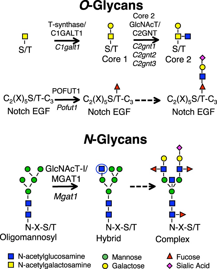
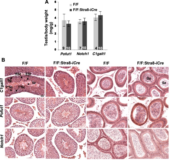
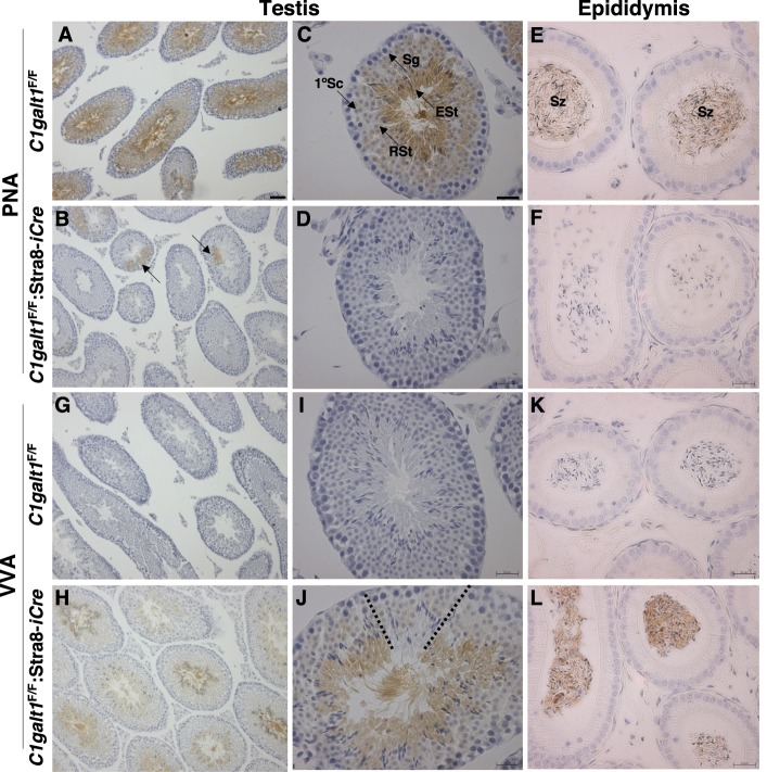
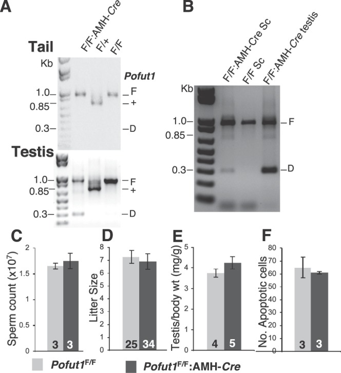
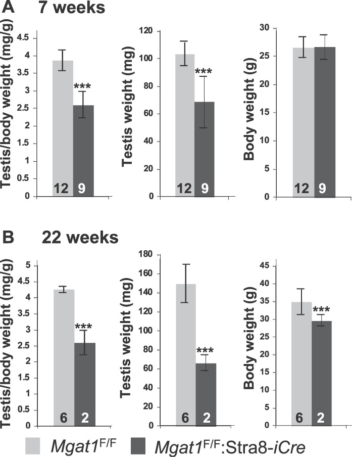
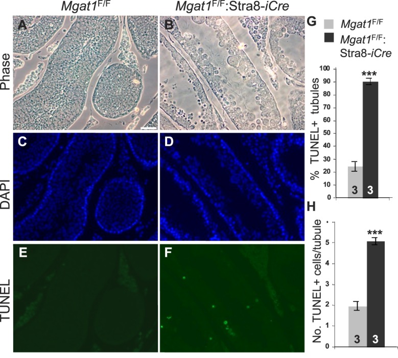
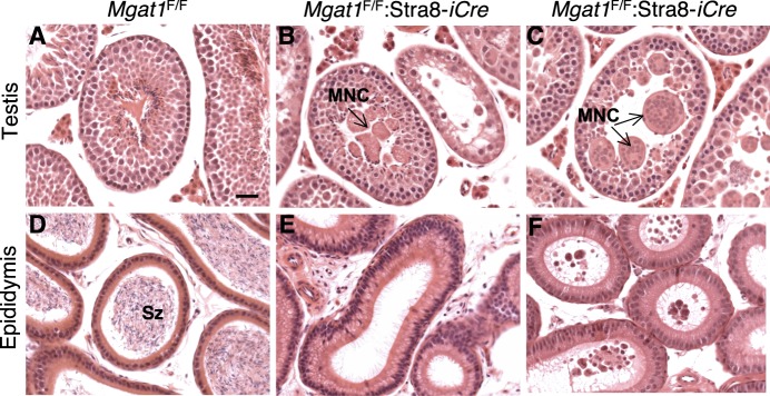
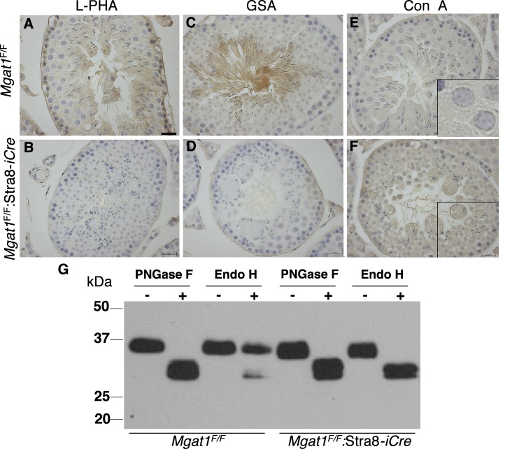
To identify roles in spermatogenesis for major subclasses of N- and O-glycans and Notch signaling, male mice carrying floxed C1galt1, Pofut1, Notch1 or Mgat1 alleles and a testis-specific Cre recombinase transgene were generated. T-synthase (C1GALT1) transfers Gal to generate core 1 and core 2 mucin O-glycans; POFUT1 transfers O-fucose to particular epidermal growth factor-like repeats and is essential for canonical Notch signaling; and MGAT1 (GlcNAcT-I) transfers GlcNAc to initiate hybrid and complex N-glycan synthesis. Cre recombinase transgenes driven by various promoters were investigated, including Stra8-iCre expressed in spermatogonia, Sycp1-Cre expressed in spermatocytes, Prm1-Cre expressed in spermatids, and AMH-Cre expressed in Sertoli cells. All Cre transgenes deleted floxed alleles, but efficiencies varied widely. Stra8-iCre was the most effective, deleting floxed Notch1 and Mgat1 alleles with 100% efficiency and floxed C1galt1 and Pofut1 alleles with ~80% efficiency, based on transmission of deleted alleles. Removal of C1galt1, Pofut1, or Notch1 in spermatogonia had no effect on testicular weight, histology, or fertility. However, males in which the synthesis of complex N-glycans was blocked by deletion of Mgat1 in spermatogonia did not produce sperm. Spermatogonia, spermatocytes, and spermatids were generated, but most spermatids formed giant multinucleated cells or symplasts, and apoptosis was increased. Therefore, although core 1 and 2 mucin O-glycans, NOTCH1, POFUT1, O-fucose glycans, and Notch signaling are dispensable, MGAT1 and complex N-glycans are essential for spermatogenesis.


Figures

FIG. 1.
FIG. 1.
Glycosyltransferase genes conditionally deleted in testis. Each reaction shown is catalyzed by the respective glycosyltransferase. Core 1 β(1,3)galactosyltransferase 1 or T-synthase/C1GALT1, encoded by the C1galt1 gene, transfers Gal to O-GalNAc on Ser/Thr residues in mucin O-glycan synthesis. Core 2 O-glycans are generated by the action of any of three core 2 β(1,6)N-acetylgucosaminyltransferases (Core 2 GlcNAcT/C2GNT) encoded by the C2gnt1, C2gnt2, or C2gnt3 genes as shown. Protein O-fucosyltransferase 1 (POFUT1), encoded by the Pofut1 gene, transfers O-fucose to EGF repeats that contain the consensus motif shown [60]; the O-fucose can be extended by the addition of GlcNAc, Gal, and sialic acid in mammalian cells [61, 62]. N-acetylglucosaminyltransferase I (GlcNAcT-I/MGAT1), encoded by the Mgat1 gene, transfers GlcNAc (circled) to a high-mannose N-glycan at N-X-S/T sequons where X is not P. This intermediate is subsequently processed and extended to form a complex N-glycan. Sugar symbols are as shown.
FIG. 2.
FIG. 2.
Deletion of C1galt1, Pofut1, or Notch1 in spermatogonia. A) Testis:body weight (mg/g) in 7-wk-old control littermates (F/F) and C1galt1F/F:Stra8-iCre, Pofut1F/F:Stra8-iCre or Notch1F/F:Stra8-iCre males. Error bars are SD. B) H&E of testis (left) and epididymal (right) sections from control (F/F) and conditional mutant males at 7 wk as marked. All panels at the same magnification; bar in A  =  10 μm. Top left panel also identifies tubule regions containing spermatogonia (Sg), 1° spermatocytes (1°Sc), 2°Sc, round spermatids (RSt), elongated spermatids (ESt), and spermatozoa (Sz). Top right panel identifies Sz in epididymal sections.
FIG. 3.
FIG. 3.
O-glycans expressed in C1galt1F/F:Stra8-iCre testis. A, B) Binding of PNA to testis sections from 7-wk-old control and C1galt1F/F:Stra8-iCre mice. Bar in A  =  50 μm. Arrows in B show small clonal regions of PNA binding in mutant testis sections. C, D) Binding of PNA to testis sections as in A, B. Bar in C = 20 μm. E, F) Binding of PNA to epididymal sections from 22-wk-old control and C1galt1F/F:Stra8-iCre mice. Top middle panel identifies tubule regions spermatogonia (Sg), 1° spermatocytes (1°Sc), round spermatids (RSt), and elongated spermatids (ESt). Top right panel identifies spermatozoa (Sz) in epididymal sections. G, H) Binding of VVA to testis sections from 7-wk-old control and C1galt1F/F:Stra8-iCre males. I, J) Binding of VVA to testis sections as in G, H. Dotted lines in J outline small clonal region in mutant testis that does not bind VVA. K, L) Binding of VVA to epididymal sections from 22-wk-old control and C1galt1F/F:Stra8-iCre mice.
FIG. 4.
FIG. 4.
Deletion of Pofut1 in Sertoli cells. A) Deletion of floxed Pofut1 alleles in testes of Pofut1F/F:AMH-Cre males. Genomic DNA from mouse tail or testes of 6- to 8-mo-old control and Pofut1F/F:AMH-Cre males was genotyped by PCR using primers PS644 and PS645 [21, 35] that amplify a 960-bp product from the floxed Pofut1 allele (F), a 700-bp product from wild-type allele (+), and a 300-bp product from the deleted allele (D). B) PCR genotyping of DNA from Sertoli cells (Sc) isolated from 3-wk-old mice and from 6-mo-old testis using primers PS644 and PS645. C) Sperm count from one caudal epididymis per mouse was examined in 6- to 8-mo-old control and Pofut1F/F:AMH-Cre mice (n = 3 each genotype). D) Litter sizes from matings of Pofut1F/F:AMH-Cre or control Pofut1F/F males with C57Bl/6 females. Eight controls had 25 litters and nine Pofut1F/F:AMH-Cre males had 34 litters. E) Testis:body weight in 6- to 8-mo-old control and Pofut1F/F:AMH-Cre males. F) Apoptosis was quantified as the average number of apoptotic cells per 100 round testis tubules. Error bars are SEM.
FIG. 5.
FIG. 5.
Deletion of Mgat1 in spermatogonia reduces testis weight. A) Testis and body weights of control and Mgat1F/F:Stra8-iCre males at 7 wk of age. B) Testis and body weights of control and Mgat1F/F:Stra8-iCre males at 22 wk of age. Error bars are SD; ***P value below 0.001 based on the Student t-test.
FIG. 6.
FIG. 6.
Apoptosis is increased in Mgat1F/F:Stra8-iCre testes. AF) Testis sections from 7-wk-old males subjected to the TUNEL assay and imaged by phase or fluorescence microscopy for apoptotic cells (green) and nuclei stained by DAPI (blue). Bar in A = 50 μm. G) The percentage of tubules containing apoptotic cells. H) The total number of apoptotic cells per apoptotic tubule. Three control and mutant testes and two independent sections for each were analyzed. Error bars are SD; ***P value below 0.001 based on the Student t-test.
FIG. 7.
FIG. 7.
Complex N-glycans are essential for spermatogenesis. AC) H&E-stained testis sections of 7-wk-old Mgat1F/F and Mgat1F/F:Stra8-iCre males. MNC labels multinucleated cells. DF) H&E-stained epididymal sections of 7-wk-old Mgat1F/F and Mgat1F/F:Stra8-iCre males (D, E) or 22 wk of age (F). Degenerate germ cells accumulated in epididymides of adult Mgat1F/F:Stra8-iCre males (F). Sz, spermatozoa. Bar in A = 10 μm.
FIG. 8.
FIG. 8.
Mgat1F/F:Stra8-iCre males do not produce complex N-glycans in germ cells. A, B) Binding of L-PHA to testis sections from 7-wk-old control and Mgat1F/F:Stra8-iCre males. C, D) Binding of GSA to testis sections as in A, B. E, F) Binding of Con A to testis sections as in A, B. Bar in A = 20 μm. G. Lysates from control and Mgat1F/F:Stra8-iCre decapsulated testes were digested with (+) PNGase F or Endo H, or incubated without (−) enzyme, and subjected to immunoblotting using anti-basigin mAb.

References

    1. Hermo L, Pelletier RM, Cyr DG, Smith CE. Surfing the wave, cycle, life history, and genes/proteins expressed by testicular germ cells. Part 1: background to spermatogenesis, spermatogonia, and spermatocytes. Microsc Res Tech 2010; 73: 241 278 - PubMed
    1. Nalam RL, Matzuk MM. Local signalling environments and human male infertility: what we can learn from mouse models. Expert Rev Mol Med 2010; 12: e15 - PMC - PubMed
    1. Yatsenko AN, Iwamori N, Iwamori T, Matzuk MM. The power of mouse genetics to study spermatogenesis. J Androl 2010; 31: 34 44 - PMC - PubMed
    1. Jones CJ, Morrison CA, Stoddart RW. Histochemical analysis of rat testicular glycoconjugates. 2. Beta-galactosyl residues in O- and N-linked glycans in seminiferous tubules. Histochem J 1992; 24: 327 336 - PubMed
    1. Jones CJ, Morrison CA, Stoddart RW. Histochemical analysis of rat testicular glycoconjugates. 1. Subsets of N-linked saccharides in seminiferous tubules. Histochem J 1992; 24: 319 326 - PubMed

Publication types