. 2012 Apr 17;109(16):6118-23.
doi: 10.1073/pnas.1200206109. Epub 2012 Apr 6.

Cryo-EM structure of a transcribing cypovirus

Affiliations

Cryo-EM structure of a transcribing cypovirus

Chongwen Yang et al. Proc Natl Acad Sci U S A. .

Abstract

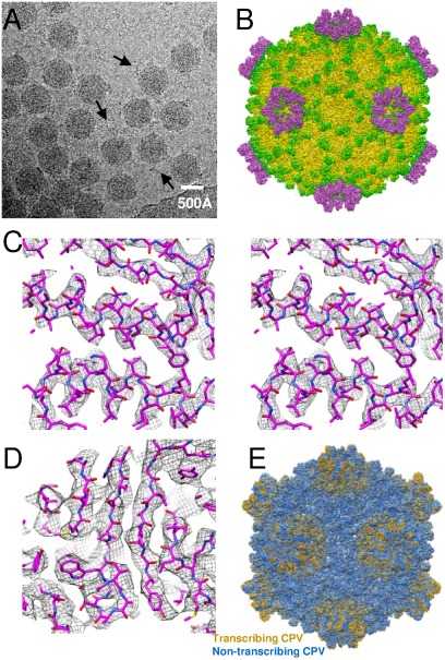
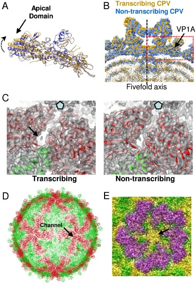
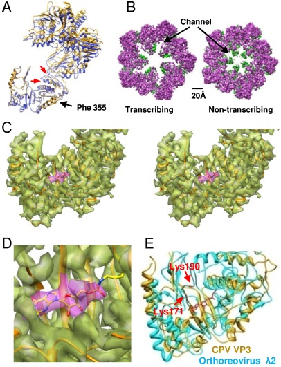
Double-stranded RNA viruses in the family Reoviridae are capable of transcribing and capping nascent mRNA within an icosahedral viral capsid that remains intact throughout repeated transcription cycles. However, how the highly coordinated mRNA transcription and capping process is facilitated by viral capsid proteins is still unknown. Cypovirus provides a good model system for studying the mRNA transcription and capping mechanism of viruses in the family Reoviridae. Here, we report a full backbone model of a transcribing cypovirus built from a near-atomic-resolution density map by cryoelectron microscopy. Compared with the structure of a nontranscribing cypovirus, the major capsid proteins of transcribing cypovirus undergo a series of conformational changes, giving rise to structural changes in the capsid shell: (i) an enlarged capsid chamber, which provides genomic RNA with more flexibility to move within the densely packed capsid, and (ii) a widened peripentonal channel in the capsid shell, which we confirmed to be a pathway for nascent mRNA. A rod-like structure attributable to a partially resolved nascent mRNA was observed in this channel. In addition, conformational change in the turret protein results in a relatively open turret at each fivefold axis. A GMP moiety, which is transferred to 5'-diphosphorylated mRNA during the mRNA capping reaction, was identified in the pocket-like guanylyltransferase domain of the turret protein.


Conflict of interest statement

The authors declare no conflict of interest.

Figures

Fig. 1.
Fig. 1.
Polyacrylamide gel electrophoresis analysis of transcription reaction mixtures with different concentrations of GTP and incubation times. All 10 RNA segments are labeled. Lane 1: transcription reaction mixture consisting of 70 mM Tris-Ac (pH 8), 10 mM MgAc2, 100 mM NaAc, 4 mM ATP, 2 mM GTP, 2 mM CTP, 2 mM UTP, 20 μCi [α-32P] UTP (specific activity 3,000 Ci/mM), 1 mM SAM, 1 U/μL RNase inhibitor and purified CPV suspension was incubated at 31 °C for 3 h. Lane 2: transcription reaction mixture in the absence of GTP (other conditions are the same as those for lane 1). Lane 3 and 4: transcription reaction mixtures in which the concentration of GTP was decreased to 0.04 mM were incubated at 31 °C for 3 h and 1 h, respectively.
Fig. 2.
Fig. 2.
Overall structure of the CPV capsid. (A) A cryo-EM image of CPV. Strand-like material was visible around many of the particles (arrows). (B) A radially colored shaded surface representation of CPV viewed along a twofold axis. (C) Wall-eye stereo view of part of the capsid shell protein α-helix density map (mesh) superimposed on the atomic model. (D) Part of the capsid shell protein β-strands density map (mesh) superimposed on the atomic model. (E) Structure of transcribing CPV (yellow) superimposed on nontranscribing CPV (blue). The spike-like structures of nontranscribing CPV were removed computationally for clarity.
Fig. 3.
Fig. 3.
Capsid shell proteins. (A) VP1A of the transcribing CPV model (yellow) superimposed on VP1A of nontranscribing CPV (blue). The direction of the apical domain tilt is indicated by an arrow. (B) A slab view of the turret of transcribing CPV (yellow) superimposed on nontranscribing CPV (blue). The structure in the red box is a copy of the VP1A of transcribing CPV (yellow) and a copy of the VP1A of nontranscribing CPV (blue), in the same orientation as that in A, indicating that the tilt of the VP1A apical domain gives rise to an enlarged capsid chamber. (C) Left: two copies of VP1A (red) and one of VP1B (blue) are superimposed on the density map of transcribing CPV. A peripentonal RNA channel and a rod-like density are indicated by an arrow. Right: two copies of VP1A (red) and a copy of VP1B (blue) are superimposed on the density map of nontranscribing CPV. These proteins have the same orientation as those on the left for comparison. The peripentonal channel on the left (transcribing) is wider than that on the right (nontranscribing). The small protrusion in the channel on the right is the side chain of VP1 Arg 653. (D) Location (arrow) of one of the peripentonal channels in the capsid shell formed by VP1A (red) and B (green). (E) The turret of transcribing CPV showing that a nodule-like structure blocks the pentameric channel at the fivefold axis.
Fig. 4.
Fig. 4.
Turret protein VP3. (A) The VP3 of transcribing CPV (yellow) superimposed on the VP3 of nontranscribing CPV (blue). Two red arrows point to conformational changes of a loop and an α-helix. (B) A comparison of the turret of transcribing and nontranscribing CPV. The color scheme is the same as that in Fig. 2B. (C) A wall-eye stereo view of the density map (transparent) of the VP3 GTase domain with its atomic model superimposed. The density map of the GMP moiety is in purple. (D) Zoom-in view of the GMP in C. (E) The CPV VP3 GTase domain is superimposed on that of orthoreovirus λ2. The locations of Lys 190 and Lys 171 in λ2 are marked. The GMP moiety in VP3 is in red.

References

    1. Mertens P. The dsRNA viruses. Virus Res. 2004;101:3–13. - PubMed
    1. Li ZL, Baker ML, Jiang W, Estes MK, Prasad BVV. Rotavirus architecture at subnanometer resolution. J Virol. 2009;83:1754–1766. - PMC - PubMed
    1. Zhang X, et al. Bluetongue virus coat protein VP2 contains sialic acid-binding domains, and VP5 resembles enveloped virus fusion proteins. Proc Natl Acad Sci USA. 2010;107:6292–6297. - PMC - PubMed
    1. Zhang X, Walker SB, Chipman PR, Nibert ML, Baker TS. Reovirus polymerase lambda 3 localized by cryo-electron microscopy of virions at a resolution of 7.6 angstrom. Nat Struct Biol. 2003;10:1011–1018. - PMC - PubMed
    1. Cheng LP, et al. Backbone model of an aquareovirus virion by cryo-electron microscopy and bioinformatics. J Mol Biol. 2010;397:852–863. - PMC - PubMed

Publication types

Associated data

LinkOut - more resources