. 2012 Jul 1;21(13):3001-12.
doi: 10.1093/hmg/dds130. Epub 2012 Apr 5.

Ube3a-ATS is an atypical RNA polymerase II transcript that represses the paternal expression of Ube3a

Affiliations

Ube3a-ATS is an atypical RNA polymerase II transcript that represses the paternal expression of Ube3a

Linyan Meng et al. Hum Mol Genet. .

Abstract

The Angelman syndrome gene, UBE3A, is subject to genomic imprinting controlled by mechanisms that are only partially understood. Its antisense transcript, UBE3A-ATS, is also imprinted and hypothesized to suppress UBE3A in cis. In this research, we showed that the mouse antisense ortholog, Ube3a-ATS, was transcribed by RNA polymerase (RNAP) II. However, unlike typical protein-coding transcripts, Ube3a-ATS was not poly-adenylated and was localized exclusively in the nucleus. It was relatively unstable with a half-life of 4 h, shorter than most protein-coding RNAs tested. To understand the role of Ube3a-ATS in vivo, a mouse model with a 0.9-kb genomic deletion over the paternal Snrpn major promoter was studied. The mice showed partial activation of paternal Ube3a, with decreased expression of Ube3a-ATS but not any imprinting defects in the Prader-Willi syndrome/Angelman syndrome region. A novel cell culture model was also generated with a transcriptional termination cassette inserted downstream of Ube3a on the paternal chromosome to reduce Ube3a-ATS transcription. In neuronally differentiated embryonic stem (ES) cells, paternal Ube3a was found to be expressed at a high level, comparable with that of the maternal allele. To further characterize the antisense RNA, a strand-specific microarray was performed. Ube3a-ATS was detectable across the entire locus of Ube3a and extended beyond the transcriptional start site of Ube3a. In summary, we conclude that Ube3a-ATS is an atypical RNAPII transcript that represses Ube3a on the paternal chromosome. These results suggest that the repression of human UBE3A-ATS may activate the expression of UBE3A from the paternal chromosome, providing a potential therapeutic strategy for patients with Angelman syndrome.


Figures

Figure 1.
Figure 1.
Ube3a-ATS is an atypical RNAPII transcript. (A) To test if Ube3a-ATS was transcribed by RNAPII, primary neuronal cultures were treated with RNAPII inhibitor α-amanitin (5 μg/ml) for 48 h. Expression levels were measured by q-PCR and normalized to 18S rRNA. (B) To test if Ube3a-ATS was poly-adenylated, mRNA purified with oligo(dT) beads was compared with total RNA by q-PCR. Pgk1 was used as the reference control and its poly-adenylation rate was set as 100%. Protein-coding genes, such as Gapdh and Actb, served as positive controls for poly-adenylated genes, and ncRNAs, such as snoRNA MBII-52 and rRNA 18S, were negative controls for non-poly-adenylated genes. (C) To test the subcellular localization of Ube3a-ATS, total RNAs were extracted from nuclear and cytoplasmic fractions of primary neuronal cultures, and the ratio of c/n was measured by q-PCR. Pgk1 was used as the reference control and its c/n ratio was set as one. Protein-coding transcripts such as Ube3a and Actb and nuclear-retained RNAs like 45S rRNA and Airn were included as controls. (D) To measure the half-life of Ube3a-ATS, primary neuronal cultures were treated with 10 μg/ml of ActD and harvested at various time points. RNA was extracted and analyzed by q-PCR. All transcripts were normalized to 18S rRNA. Transcripts are indicated as follows: dashed-dotted line for c-Myc, dashed lines for Ube3a, Actb and Airn, solid lines for Ube3a-ATS. All the experiments were repeated at least two times with a representative result showing here. Error bars represent the standard error of means of three technical replicates.
Figure 2.
Figure 2.
Both 0.9- and 4.8-kb deletions at the Snrpn promoter activate the paternal expression of Ube3a. (A) The PWS/AS imprinted region is shown with paternally expressed genes highlighted in blue, maternally expressed genes in red and silenced genes in gray (not drawn to scale). Ube3a-ATS is proposed to be processed from the same precursor encoding Snrpn, which has multiple upstream promoters. The major promoter is located within PWS-IC. Regions deleted in each mutant allele are shown by the green bars: del4.8 covers most of the PWS-IC; del0.9 covers the Snrpn major promoter and a small part of PWS-IC; dels-u is a deletion from Snrpn to Ube3a. The open circle represents unmethylated status and the closed circle represents methylated status. Pat, paternal; Mat, maternal. (B) Bisulfite sequencing of Snrpn intron1 downstream of the 0.9-kb deletion region was performed with DNA extracted from the cerebral cortex of WT and del+/0.9 mice at P14. Mice analyzed were the F1 generation of a cross between male C57BL/6J del+/0.9 mice and female CAST.chr7 WT mice. Clones representing the maternal allele were distinguished by the conversion of a CpG dinucleotide (CG > AA). A total of 13 CpG dinucleotides were analyzed with open circles for unmethylated CpG and closed circle for methylated CpG. Ten random clones from each allele were shown with each line representing sequences from one clone. (C) The expression pattern of selected genes located in the PWS/AS region were compared in newborn mouse brains of dels-u/+, dels-u/0.9, dels-u/4.8 and del+/s-u mutants by q-PCR. All transcripts were normalized to the internal control of Pgk1. For better illustration, del+/s-u was used as the reference when the Ube3a level was plotted, and dels-u/+ was the reference when plotting the rest of the genes. Three biological replicates were performed with one representative result shown here. Error bars represent the standard error of means from three technical repeats. (D) Western blot was performed with proteins extracted from newborn mouse brains of dels-u/+, dels-u/0.9, dels-u/4.8 and del+/s-u mutants. β-Tubulin was used as the loading control. (E) Brain sections from 1-month-old mice were immunostained with anti-Ube3a (green) and anti-NeuN (red). Expression of paternal Ube3a was detected in cortical and hippocampal neurons in the dels-u/0.9 and dels-u/4.8 mutants. Ctx, cerebral cortex; WT, wild-type.
Figure 3.
Figure 3.
Early termination of Ube3a-ATS activates paternal Ube3a in cultured differentiated neurons. (A) Male Ube3a+/YFP mice were crossed with female WT mice to generate primary ES cells of Ube3a+/YFP. The ES cells were then electroporated with a targeting vector, selected with neomycin and screened for positive clones by Southern blot. Clones with stop allele inserted in cis or in trans of the Ube3aYFP allele were also identified by Southern blot. The cis and trans clones (Ube3a+/YFP;stop and Ube3aYFP/stop), together with control clones (negative control of Ube3a+/YFP and positive control of Ube3aYFP/+), were differentiated into neuronal linage and harvested for RNA and protein analysis. (B) The transcription termination cassette was engineered into the genome 11.9-kb downstream of Ube3a. The cassette consists of a pair of loxP sites (triangles), a splicing donor (black box), a triple polyA signal derived from SV40 (white boxes) and FRT-flanked PGK-neomycin (white box flanked with gray boxes). (C) Using q-PCR, the levels of Ube3aYFP and Ube3a-ATSYFP were determined in differentiated neuronal cells of Ube3aYFP/+, Ube3a+/YFP, Ube3astop/YFP and Ube3a+/YFP;stop genotypes. Both transcripts were normalized to the internal control of Pgk1. (D) Differentiated neuronal cells were immunostained with anti-YFP and neuronal marker anti-Map2. The top panels show anti-YFP signal only and the bottom panels are merge of anti-YFP (green) and anti-Map2 (red). (E) YFP intensity was quantified in Map2 positive cells of four different clones by ImageJ. Error bars represent the standard error of means. Statistical analysis was performed with Student's t-test comparing the sample with the control of Ube3a+/YFP. ns, not significant; *P < 0.01.
Figure 4.
Figure 4.
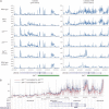
Differential expression of Ube3a and Ube3a-ATS is detected by the strand-specific microarray. (A) Total RNAs or polyA+ RNAs extracted from newborn mouse brains with various genotypes were reverse-transcribed, labeled and hybridized to the custom-designed strand-specific microarray. The microarray includes probes from both plus and minus strands of the Ube3a region, which hybridized to Ube3a and Ube3a-ATS cDNA, respectively. Normalized signal intensity (y-axis) was plotted against genomic coordinates (x-axis, NCBI37/mm9 build). The genomic segment deleted in dels-u is marked by the green bar. Noisy peaks to the left of the deletion boundary in some of the samples are probably artifacts arising from the ectopic expression of the selection marker inserted in the deletion region. (B) To reduce the background noise, the relative intensity of Ube3a-ATS was calculated as the difference between dels-u/+ and dels-u/0.9 or dels-u/4.8. The light lines represent the relative intensity and the dark lines are the moving average of 20 probes.

References

    1. Edwards C.A., Ferguson-Smith A.C. Mechanisms regulating imprinted genes in clusters. Curr. Opin. Cell Biol. 2007;19:281–289. doi:10.1016/j.ceb.2007.04.013. - DOI - PubMed
    1. Horsthemke B. Mechanisms of imprint dysregulation. Am. J. Med. Genet. C Semin. Med. Genet. 2010;154C:321–328. doi:10.1002/ajmg.c.30269. - DOI - PubMed
    1. Buiting K. Prader-Willi syndrome and Angelman syndrome. Am. J. Med. Genet. C Semin. Med. Genet. 2010;154C:365–376. doi:10.1002/ajmg.c.30273. - DOI - PubMed
    1. Horsthemke B., Wagstaff J. Mechanisms of imprinting of the Prader-Willi/Angelman region. Am. J. Med. Genet. A. 2008;146A:2041–2052. doi:10.1002/ajmg.a.32364. - DOI - PubMed
    1. Kantor B., Makedonski K., Green-Finberg Y., Shemer R., Razin A. Control elements within the PWS/AS imprinting box and their function in the imprinting process. Hum. Mol. Genet. 2004;13:751–762. doi:10.1093/hmg/ddh085. - DOI - PubMed

Publication types

MeSH terms