. 2012 Apr 3;3(2):e00282-11.
doi: 10.1128/mBio.00282-11. Print 2012.

Characterization of cleavage events in the multifunctional cilium adhesin Mhp684 (P146) reveals a mechanism by which Mycoplasma hyopneumoniae regulates surface topography

Affiliations

Characterization of cleavage events in the multifunctional cilium adhesin Mhp684 (P146) reveals a mechanism by which Mycoplasma hyopneumoniae regulates surface topography

Daniel R Bogema et al. mBio. .

Abstract

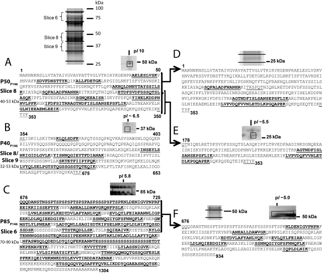
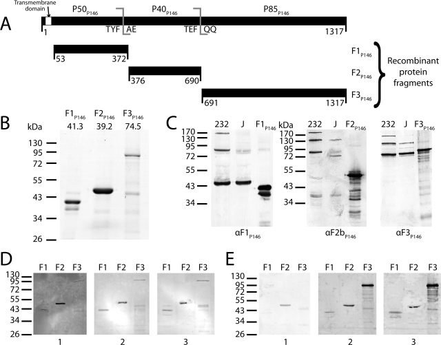
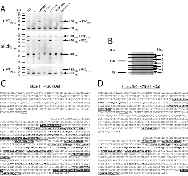
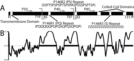
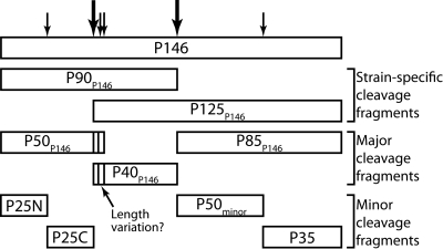
Mycoplasma hyopneumoniae causes enormous economic losses to swine production worldwide by colonizing the ciliated epithelium in the porcine respiratory tract, resulting in widespread damage to the mucociliary escalator, prolonged inflammation, reduced weight gain, and secondary infections. Protein Mhp684 (P146) comprises 1,317 amino acids, and while the N-terminal 400 residues display significant sequence identity to the archetype cilium adhesin P97, the remainder of the molecule is novel and displays unusual motifs. Proteome analysis shows that P146 preprotein is endogenously cleaved into three major fragments identified here as P50(P146), P40(P146), and P85(P146) that reside on the cell surface. Liquid chromatography with tandem mass spectrometry (LC-MS/MS) identified a semitryptic peptide that delineated a major cleavage site in Mhp684. Cleavage occurred at the phenylalanine residue within sequence (672)ATEF↓QQ(677), consistent with a cleavage motif resembling S/T-X-F↓X-D/E recently identified in Mhp683 and other P97/P102 family members. Biotinylated surface proteins recovered by avidin chromatography and separated by two-dimensional gel electrophoresis (2-D GE) showed that more-extensive endoproteolytic cleavage of P146 occurs. Recombinant fragments F1(P146)-F3(P146) that mimic P50(P146), P40(P146), and P85(P146) were constructed and shown to bind porcine epithelial cilia and biotinylated heparin with physiologically relevant affinity. Recombinant versions of F3(P146) generated from M. hyopneumoniae strain J and 232 sequences strongly bind porcine plasminogen, and the removal of their respective C-terminal lysine and arginine residues significantly reduces this interaction. These data reveal that P146 is an extensively processed, multifunctional adhesin of M. hyopneumoniae. Extensive cleavage coupled with variable cleavage efficiency provides a mechanism by which M. hyopneumoniae regulates protein topography.

Importance: Vaccines used to control Mycoplasma hyopneumoniae infection provide only partial protection. Proteins of the P97/P102 families are highly expressed, functionally redundant molecules that are substrates of endoproteases that generate multifunctional adhesin fragments on the cell surface. We show that P146 displays a chimeric structure consisting of an N terminus, which shares sequence identity with P97, and novel central and C-terminal regions. P146 is endoproteolytically processed at multiple sites, generating at least nine fragments on the surface of M. hyopneumoniae. Dominant cleavage events occurred at S/T-X-F↓X-D/E-like sites generating P50(P146), P40(P146), and P85(P146). Recombinant proteins designed to mimic the major cleavage fragments bind porcine cilia, heparin, and plasminogen. P146 undergoes endoproteolytic processing events at multiple sites and with differential processing efficiency, generating combinatorial diversity on the surface of M. hyopneumoniae.


Figures

FIG 1
FIG 1
Mass spectrometry analysis of P146. M. hyopneumoniae strain J proteins were examined using the following two methods. In method 1, lysates were subjected to 1-D SDS-PAGE, the gels were sectioned into pieces representing 14 molecular mass ranges, and the proteins in each gel slice were examined using LC-MS/MS. In method 2, surface proteins were labeled by surface biotinylation and enriched by affinity chromatography of the cell lysate. Purified biotinylated proteins were separated by either 1-D SDS-PAGE or 2-D GE and identified by LC-MS/MS. Peptides identified by LC-MS/MS are shown in bold type and underlined. The numbers above or below the sequence refer to amino acid positions. (A to C) Peptides unique to three regions of the P146 sequence were identified in both experiments. Terminal sequences matching an M. hyopneumoniae cleavage motif defined previously are underlined but not bold. (A) Fragment P50P146 and peptides unique to the N-terminal region of P146. (B) Fragment P40P146 and peptides unique to the central region of P146. (C) Fragment P85P146 and peptides unique to the C-terminal region of P146. The N terminus of P85P146 was identified from a semitryptic peptide. (D to F) Additional proteins were identified by method 2 at masses significantly lower than those identified by method 1. Terminal sequences matching or almost matching an M. hyopneumoniae cleavage motif defined previously (22) are underlined but not bold. (D) Peptides unique to P50P146 were identified in a 1-D SDS-polyacrylamide gel slice containing proteins migrating at ~25 kDa. (E) Peptides unique to one-half of P50P146 identified in a 2-D GE protein spot migrating at 25 kDa and pI of ~5.5. (F) Peptides unique to P85P146 identified in 2-D GE protein spots migrating at 50 kDa and pI of ~5.0.
FIG 2
FIG 2
Cloning of p146 and immunoblot analysis of its cleavage fragments. (A) p146 (3954 bp encoding 1317 residues) was cloned from M. hyopneumoniae strain 232 homolog mhp684 in three fragments (F1P146/232 to F3P146/232) closely matching native cleavage as observed by mass spectrometry. A hydrophobic transmembrane domain predicted by TMHMM (53 residues) was removed from the N terminus of F1P146/232. All in-frame TGA codons were mutated to TGG using overlap extension PCR. (B) Coomassie blue-stained SDS-PAGE of the three P146 recombinant proteins. The predicted mass (in kilodaltons) is shown above each gel lane. Recombinant protein F2P146/232 runs at a higher molecular mass than predicted. (C) Immunoblots of recombinant proteins (F1P146/232 to F3P146/232) and whole-cell lysates from M. hyopneumoniae strains 232 and J, probed with anti-F1P146 (αF1P146) and anti-F3P146 (αF3P146) sera. Sera labeled αF1P146 and αF3P146 were extracted from New Zealand White rabbits challenged with recombinants F1P146/232 and F3P146/232; anti-F2bP146 (αF2bP146) serum was generated in the same manner from a recombinant protein representing residues 586 to 690 of P146. Minor bands present in recombinant protein lanes (F1P146/232 to F3P146/232) may be either products of recombinant protein degradation or truncated proteins resulting from mRNA instability. (D) Immunoblots of recombinant proteins (F1P146/232 to F3P146/232) probed with porcine serum sourced from a high-health-status herd (blot 1) and sera from convalescent pigs (blots 2 and 3). (E) Immunoblots of recombinant proteins (F1P146/232 to F3P146/232) probed with sera obtained from a single pig prior to treatment (blot 1), after vaccination with the commercial bacterin vaccine Suvaxyn (blot 2), or 6 weeks after challenge with M. hyopneumoniae strain Hillcrest (blot 3).
FIG 3
FIG 3
P146 is processed variably in M. hyopneumoniae laboratory strains and field isolates. (A) Immunoblots of whole-cell lysates of different M. hyopneumoniae strains and field isolates. The blots were separately probed with anti-F1P146 (αF1P146), anti-F2bP146 (αF2bP146), and anti-F3P146 (αF3P146) sera in order to assess the consistency of protein expression and processing. Cleavage fragments equivalent to P50P146, P40P146, and P85P146 are conserved across all isolates in this study, but some also show distinct bands at higher masses that correspond to fragments that would result from inefficient cleavage. (B) 1-D SDS-polyacrylamide gel (molecular mass region ~75 to 125 kDa) of an M. hyopneumoniae strain 232 cell lysate was sectioned into 6 slices, and each slice was examined using LC-MS/MS. (C) Slice 1 (~110 to 125 kDa) was found to contain peptides unique to regions of P146 corresponding to P40P146 and P85P146 fragments and matched a band found at a similar mass in strain 232 immunoblots. (D) Slices 5 and 6 (~75 to 85 kDa) were found to contain peptides unique to all major fragments of P146, suggesting that a cleavage event separated the protein into two separate ~80-kDa fragments.
FIG 4
FIG 4
In silico analysis of P146. (A) Sequence features of P146. The TMHMM algorithm predicts that P146 contains a transmembrane domain (P = 0.999). P146 contains three defined repeat regions: P146R1 (PQ), P146R2 (PS), and P146R3 (S). Multiple coiled-coil prediction algorithms identified two putative coiled-coils flanking the P146R3 repeat region in P85P146. The experimentally determined cleavage site (ATEFQQ) and predicted site (KTYFAE) are also shown. (B) PONDR VSL1 analysis of P146 232 homolog (Mhp684). The regions above the line at 0.5 denote regions predicted to be structurally disordered within P146. The thick black bars denote disordered regions spanning 40 or more amino acids. P146 is predicted to contain five regions of significant disorder, two corresponding with the experimental and predicted cleavage sites. The remaining disordered regions correspond to putative coiled-coil domains, the C-terminal region, and the P146R3 repeat region.
FIG 5
FIG 5
Summary of P146 cleavage. Cleavage fragments can be split into three groups as shown. Strain-specific cleavage fragments are prevalent fragments in selected strains (e.g., strain 232). Major cleavage fragments P50P146, P40P146, and P85P146 are abundant in all strains. Minor cleavage fragments are present in small amounts and are detected only by enrichment of surface proteins.

References

    1. Browning GF, Marenda MS, Noormohammadi AH, Markham PF. 2011. The central role of lipoproteins in the pathogenesis of mycoplasmoses. Vet. Microbiol. 153:44–50 - PubMed
    1. Rosengarten R, Wise KS. 1991. The Vlp system of Mycoplasma hyorhinis: combinatorial expression of distinct size variant lipoproteins generating high-frequency surface antigenic variation. J. Bacteriol. 173:4782–4793 - PMC - PubMed
    1. Citti C, Watson-McKown R, Droesse M, Wise KS. 2000. Gene families encoding phase- and size-variable surface lipoproteins of Mycoplasma hyorhinis. J. Bacteriol. 182:1356–1363 - PMC - PubMed
    1. Noormohammadi AH, et al. 1997. Mycoplasma synoviae has two distinct phase-variable major membrane antigens, one of which is a putative hemagglutinin. Infect. Immun. 65:2542–2547 - PMC - PubMed
    1. Liu W, et al. 2011. Complete genome sequence of Mycoplasma hyopneumoniae strain 168. J. Bacteriol. 193:1016–1017 - PMC - PubMed

Publication types