. 2012 Jun;32(12):2196-202.
doi: 10.1128/MCB.06294-11. Epub 2012 Apr 9.

Protection from bacterial-toxin-induced apoptosis in macrophages requires the lipogenic transcription factor sterol regulatory element binding protein 1a

Affiliations

Protection from bacterial-toxin-induced apoptosis in macrophages requires the lipogenic transcription factor sterol regulatory element binding protein 1a

Seung-Soon Im et al. Mol Cell Biol. 2012 Jun.

Abstract

Sterol regulatory element binding protein (SREBP) transcription factors activate genes of lipid metabolism, but recent studies indicate they also activate genes involved in other physiologic processes, suggesting that SREPBs have evolved to connect lipid metabolism with diverse physiologic responses. There are three major mammalian SREBPs, and the 1a isoform is specifically expressed at very high levels in macrophages, where a recent study showed that it couples lipid synthesis to the proinflammatory phase of the innate immune response. In the present study, we show that loss of SREBP-1a also results in an increase in apoptosis after exposure to bacterial pore-forming toxins and we show this is a result of a selective reduction in the expression of the gene coding for the antiapoptotic factor apoptosis inhibitor 6 (Api6). Additional studies demonstrate that SREBP-1a specifically activates the Api6 gene through a binding site in its proximal promoter, thus establishing the Api6 gene as a newly identified SREBP-1a target gene.


Figures

Fig 1
Fig 1
Examination of S. aureus alpha-toxin-induced apoptosis. (A) Analysis of alpha-toxin-induced cell death by flow cytometry. Completely differentiated bone marrow-derived macrophages (BMDMs) (4 × 10⁵ cells/well) were plated in a 6-well plate. WT and SREBP-1aDF macrophages were treated with 5 ng/μl alpha-toxin for 24 h with 5% CO2 at 37°C; representative dot plots of annexin V (AV) and propidium iodide (PI) staining are shown. (B) Histograms from flow cytometry analyses for apoptosis by alpha-toxin measured using annexin V staining in BMDMs. BMDMs from WT or SREBP-1aDF macrophages were treated for 24 h with alpha-toxin, cells were stained with annexin V and propidium iodide, and apoptosis was quantified as described in Materials and Methods. WT versus SREBP-1aDF, P = 0.0003. Each point represents data from a separate batch of isolated macrophages. (C) Time course for apoptosis induced by toxin treatment. Macrophages were harvested at the indicated time after toxin treatment, and the percentage of apoptosis (annexin V-positive cells) was measured. Results were from at least three different experiments (n = 3 to 5 mice per group).
Fig 2
Fig 2
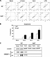
Apoptosis rates of BMDMs with streptolysin O (SLO) and LPS treatment detected by flow cytometry. BMDMs were treated with 4 μg/ml streptolysin O and 100 ng/ml LPS for 24 h, respectively. Data are shown as means ± standard deviations (n = 3). P < 0.05 was regarded as statistically significant. (A) Annexin V-FITC and PI staining for apoptosis detected by flow cytometry. (B) The graph shows the apoptosis rate of panel A. (C) Immunoblot analysis of SREBP-1 in WT and SREBP-1aDF macrophages. BMDMs were treated with 5 ng/μl of alpha-toxin and 4 μg/ml streptolysin O for 24 h and were harvested for membrane and nuclear fractions. Aliquots of 10 μg of nuclear protein extracts were subjected to SDS-PAGE, and immunoblot analysis was carried out with anti-SREBP-1. “P” and “M” denote the positions of the membrane-bound precursor and soluble, mature SREBP transcription factor, respectively.
Fig 3
Fig 3
Lipogenic gene expression levels and apoptosis by alpha-toxin treatment in macrophages. (A to E) qPCR for specific SREBPs, ACC1, and fatty acid synthase (FAS). BMDMs from WT or SREBP-1aDF mice were treated with 5 ng/μl of alpha-toxin for 24 h, and RNA was prepared. mRNA expression was measured by qPCR. Expression levels were normalized to L32 internal control gene expression individually. Bars represent the standard deviations. Representative data from three independent experiments are shown.
Fig 4
Fig 4
Api6 gene expression and apoptosis by overexpression of SREBP-1a in SREBP-1aDF macrophages. (A) mRNA expression of the Api6 gene. RNA was isolated from BMDMs of WT and SREBP-1aDF mice after alpha-toxin treatment for 24 h, and Api6 mRNA expression was measured by qPCR. (B) Effect of SREBP-1a overexpression in SREBP-1aDF macrophages. BMDMs from SREBP-1aDF mice were infected with 6 × 10⁹ PFU (MOI, 10) of adenovirus expressing GFP (Ad-GFP) or adenovirus expressing SRBEP-1a (Ad-SR1a) for 24 h and treated with alpha-toxin for a further 24 h. (C) Apoptosis was monitored by flow cytometry. Data are presented as percentages of apoptosis ± standard errors of the mean (SEM) (n = 3 mice per group). (D) Western blot for SREBP-1 by overexpression of SREBP-1a. BMDMs from WT and SREBP-1aDF mice were overexpressed with adenovirus expressing GFP and SREBP-1a for 48 h, and then membrane and nuclear protein extracts were subjected to immunoblot analysis for SREBP-1a and, as a loading control, β-actin. Precursor (p) and mature (m) forms of SREBP are noted.
Fig 5
Fig 5
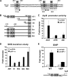
Api6 promoter activity and chromatin immunoprecipitation (ChIP) assay. (A) Comparison of SREBP binding element among humans and mice. A predicted SREBP binding element is shaded on the Api6 promoter between bp −476 and −470. (B) Serial deletion constructs of the mouse Api6 promoter. The mouse Api6 promoter was amplified from mouse genomic DNA and inserted into a luciferase (Luc) reporter vector. The luciferase activity was normalized by β-galactosidase driven by a CMV promoter. (C) Deletion constructs for the Api6 promoter were analyzed for promoter activity. (D) Chromatin from BMDMs was analyzed for recruitment of SREBP-1 to the SRE region of the Api6 promoter by ChIP as described in Materials and Methods. The quantity of DNA in the precipitation with SREBP-1 antibody was normalized to input chromatin and plotted relative to the IgG background.
Fig 6
Fig 6
Api6 mRNA level and apoptosis level after overexpression of Api6 in SREBP-1aDF macrophages. (A) Overexpression of the Api6 mRNA level after Api6 was transfected into macrophages was confirmed by qPCR. RNA was isolated from BMDMs of WT and SREBP-1aDF mice, and Api6 mRNA expression was measured by qPCR. (B) Apoptosis (annexin V-positive cells) was measured by flow cytometry. One hundred nanograms of GFP or Api6 plasmid was transfected by electroporation, and the amounts of annexin V-positive macrophages were measured by flow cytometry after 24 h. (C and D) qPCR analysis of FAS and ACC1 from BMDMs overexpressing Api6 stimulated for 24 h with alpha-toxin. Results are shown as the mean of triplicate samples ± SEM relative to unstimulated WT samples (n = 3 mice per group).
Fig 7
Fig 7
Scheme of the antiapoptotic role of SREBP-1a through the Api6 gene in macrophages. A model for how SREBP-1a regulates bacterial-toxin-induced apoptosis of the macrophage through direct activation of the antiapoptotic factor Api6 gene is shown. Our results suggest that bacterial alpha-toxin-induced SREBP-1a activates not only lipogenesis but also the antiapoptotic factor Api6 gene to limit apoptosis following bacterial infection of macrophages.

References

    1. Aderem A, Underhill DM. 1999. Mechanisms of phagocytosis in macrophages. Annu. Rev. Immunol. 17: 593– 623 - PubMed
    1. Bhakdi S, Tranum-Jensen J. 1991. Alpha-toxin of Staphylococcus aureus. Microbiol. Rev. 55: 733– 751 - PMC - PubMed
    1. Earnshaw WC, Martins LM, Kaufmann SH. 1999. Mammalian caspases: structure, activation, substrates, and functions during apoptosis. Annu. Rev. Biochem. 68: 383– 424 - PubMed
    1. Gagnon E, et al. 2002. Endoplasmic reticulum-mediated phagocytosis is a mechanism of entry into macrophages. Cell 110: 119– 131 - PubMed
    1. Gurcel L, Abrami L, Girardin S, Tschopp J, van der Goot FG. 2006. Caspase-1 activation of lipid metabolic pathways in response to bacterial pore-forming toxins promotes cell survival. Cell 126: 1135– 1145 - PubMed

Publication types

MeSH terms

Substances

LinkOut - more resources