. 2012 Jun;32(12):2268-78.
doi: 10.1128/MCB.00063-12. Epub 2012 Apr 9.

PERK utilizes intrinsic lipid kinase activity to generate phosphatidic acid, mediate Akt activation, and promote adipocyte differentiation

Affiliations

PERK utilizes intrinsic lipid kinase activity to generate phosphatidic acid, mediate Akt activation, and promote adipocyte differentiation

Ekaterina Bobrovnikova-Marjon et al. Mol Cell Biol. 2012 Jun.

Abstract

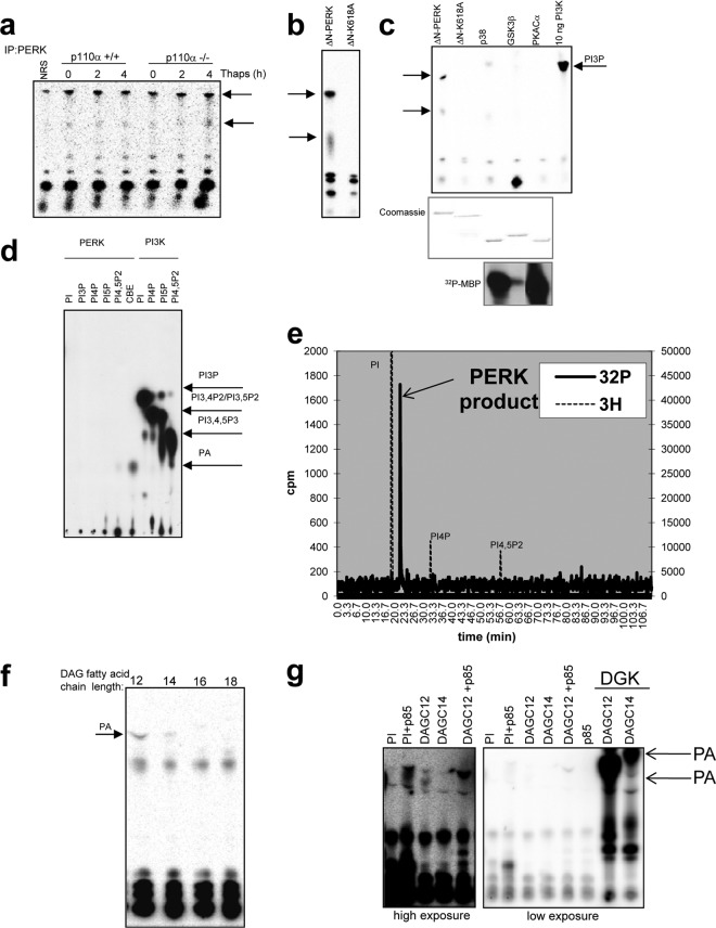
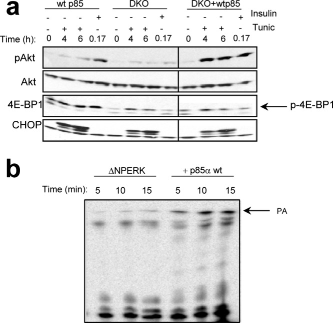
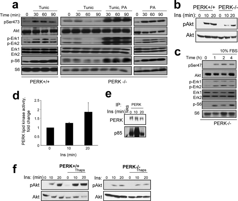
The endoplasmic reticulum (ER) resident PKR-like kinase (PERK) is necessary for Akt activation in response to ER stress. We demonstrate that PERK harbors intrinsic lipid kinase, favoring diacylglycerol (DAG) as a substrate and generating phosphatidic acid (PA). This activity of PERK correlates with activation of mTOR and phosphorylation of Akt on Ser473. PERK lipid kinase activity is regulated in a phosphatidylinositol 3-kinase (PI3K) p85α-dependent manner. Moreover, PERK activity is essential during adipocyte differentiation. Because PA and Akt regulate many cellular functions, including cellular survival, proliferation, migratory responses, and metabolic adaptation, our findings suggest that PERK has a more extensive role in insulin signaling, insulin resistance, obesity, and tumorigenesis than previously thought.


Figures

Fig 1
Fig 1
Activation of Akt during ER stress is independent of eIF2α phosphorylation and is correlated with activation of mTOR. (a) Western blot analysis for phospho-Ser473 Akt (pAkt) in eIF2α wild-type (wt) and eIF2αS51A knock-in fibroblasts following exposure of cells to tunicamycin (Tunic). (b) Activation of Akt in response to ER stress in NIH 3T3 cells coadministered with 10 nM wortmannin, 10 μM PIK-294, or 10 μM IC-87114 to inhibit endogenous PI3K activity. (c) Activation of Akt during ER stress in H1299 cells overexpressing p85α wt and p110α, p85α wt, and dominant negative (DN) p110α. (d) PERK+/+ or PERK−/− cells were treated with tunicamycin for the times indicated, and lysates were resolved by SDS-PAGE or utilized to pull down eIF4E complex with 7-Met-GTP-Sepharose. Arrows indicate relative positions of phosphorylated 4E-PB1 and CHOP.
Fig 2
Fig 2
The ER stress sensor PERK demonstrates lipid kinase activity in vitro and in vivo. (a) PERK was immunopurified from p110+/+ or p110α−/− MEFs, and lipid kinase activity was determined in an in vitro assay with phosphatidylinositol as a substrate. NRS, nonspecific rabbit serum. Thaps, thapsigargin. (b) In vitro lipid kinase assay with phosphatidylinositol as a substrate and recombinant catalytic domain of PERK (GST-ΔNPERK) or kinase-dead PERK (K618A). (c) In vitro lipid kinase assay with phosphatidylinositol as a substrate and recombinant catalytic domain of PERK (GST-ΔNPERK), kinase-dead PERK (K618A), p38, GSK3β, PKA C (500 ng of each kinase), and PI3K p85/p110 complex. (d) In vitro lipid kinase assay with phosphatidylinositol as a substrate and recombinant catalytic domain of PERK (GST-ΔNPERK) or recombinant PI3K and various phosphoinositide substrates. CBE, crude brain extract. Products were resolved by chromatography with propanol and acetic acid as the mobile phase. (e) The radioactive spot from the TLC plate in panel d (with CBE as a substrate) was excised, and PERK product was deacylated and then resolved by HPLC with 3H-labeled PI, PI4P, and PI4,5P2 as standards. (f) In vitro lipid kinase assay with diacylglycerol (DAG) as a substrate and recombinant catalytic domain of PERK (GST-ΔNPERK). DAGC12, 1,2-dilauroyl-sn-glycerol. (g) In vitro lipid kinase assay with phosphatidylinositol (PI) or diacylglycerol (DAG) as a substrate and the recombinant catalytic domain of PERK in the presence or absence of recombinant PI3K p85α or with recombinant diacylglycerol kinase (DGK). DAGC14, 1,2-dimyristoyl-sn-glycerol; DAGC16, 1,2-dipalmitoyl-sn-glycerol; DAGC18, 1,2-distearoyl-sn-glycerol.
Fig 3
Fig 3
PERK-dependent generation of PA in vivo. (a) Total phosphatidic acid (PA) content in lysates from PERK+/+ and PERK−/− MEFs that were treated with 500 nM thapsigargin (Thaps) for the times indicated. (b) Total phosphatidic acid (PA) content in lysates from wild-type (wt) and eIF2αS51A knock-in cells treated with 500 nM thapsigargin for the times indicated. (c) PA formation in MEFs treated with 500 nM thapsigargin and either DGK inhibitor R59949 (20 μM) or PLD inhibitor FIPI (1.5 μM). (d) Total PA content in lysates from wild-type MEFs treated with 10 nM wortmannin (Wort), 25 nM rapamycin-specific inhibitor against mTOR (Rap), and 1 mM MK-2206-specific inhibitor against Akt. Thaps (500 nM) was used to induce ER stress. (e) FRET-based PA biosensors were used to measure PERK-dependent PA production in live cells: wild-type and PERK−/− MEFs were transfected with Pii-DK (wild type) or Pii-DK-9A (PA binding domain mutant) constructs. CFP and FRET (red, high FRET; blue, no/low FRET; see scale on right) images were obtained. Schematic representation of the FRET biosensor is provided. FRET signal decreases when the wild-type biosensor recognizes PA. (f) Values for FRET and CFP levels were obtained using fluorescence microscopy before and after treatment with 500 nM thapsigargin. FRET/CFP ratios were calculated for both wild-type (WT) and mutant (MUT) biosensors. (g) Fold change in PA measured 4 h after thapsigargin treatment (500 nM). Significant changes were observed between PERK+/+ MEFs transfected with WT versus MUT biosensor and PERK−/− cells expressing WT versus MUT biosensor (*, P < 0.001, determined by Student t test).
Fig 4
Fig 4
PERK directly binds the regulatory subunit of PI3K in vitro and in vivo. (a) Activation of Akt in response to ER stress or in p85α/β double-knockout MEFs. (b) In vitro binding assay using GST-ΔNPERK and in vitro transcribed and translated p85α. (c) Coimmunoprecipitation of Myc-tagged PERKK618A and HA-p85α/FLAG-p110 in 293T cells. The diagram depicts deletion mutants of PERK cytosolic kinase domain. SP, signal peptide; TM, transmembrane domain. (d) Control for the experiment shown in panel c. Immunoprecipitation and Western blot with anti-myc antibody are shown. (e) Immunoprecipitation and Western blot for PERK and p85α from PERK+/+ MEFs treated with 50 nM thapsigargin (Thaps) for 2 or 4 h or stimulated with 100 nM insulin (Ins) for 15 or 30 min.
Fig 5
Fig 5
p85 increases PERK lipid kinase activity and Akt activation. (a) Activation of Akt in response to ER stress in wild-type, p85/p85 double-knockout (DKO), or DKO MEFs reconstituted with wild-type p85α (+wt p85). Phosphorylated p-4EBP1 is indicated by the arrow. (b) In vitro lipid kinase with DAGC12 as a substrate and recombinant catalytic domain of PERK (GST-ΔNPERK) alone or in the presence of recombinant p85α wild type. The intensity of phosphorylated lipid product signal was measured using phosphor imaging screen and STORM scanner (graph shown).
Fig 6
Fig 6
PERK promotes mitogenic signaling. (a) PI3K-mTOR-Akt and Ras-MEK-Erk1/2 pathway activation downstream of PERK was measured in PERK+/+/PERK−/− MEFs treated with tunicamycin (Tunic) and assessed by Western blotting using phospho-Ser473 Akt (pSer473), phospho-Erk1/2 (p-Erk1/2), and phospho-S6 kinase (p-S6) antibodies. PA was added to PERK−/− MEF cell culture media where indicated at 100 μM. (b) PERK+/+ and PERK−/− cells were serum starved overnight and then treated with 100 nM insulin for 10 or 20 min. Akt activation and levels were subsequently determined. (c) The indicated cells were serum deprived for 16 h, followed by addition of medium containing 10% FBS where indicated. Pathway activation was monitored by Western blot analysis with phospho-specific antibodies as indicated. (d) Lipid kinase activity in PERK immune complexes. The graph represents average activity, and the error bars are standard deviations from results of three independent experiments. (e) PERK was immunopurified from cells treated with 100 nM insulin, and the levels of associated p85 were assessed by Western blot analysis. (f) PERK+/+ or PERK−/− cells were serum starved overnight and then left untreated or pretreated with 500 nM thapsigargin for 4 h followed by 100 nM insulin as indicated. Lysates were probed for phospho-Ser473 Akt and total Akt levels. NRS, nonspecific rabbit serum.
Fig 7
Fig 7
Role of PERK lipid kinase activity in adipocyte differentiation in vitro. (a) Levels of phospho-Ser473 Akt in adipocytes differentiating from PERK+/+ or PERK−/− MEFs. (b) Oil Red O staining of adipocytes differentiating from PERK+/+ or PERK−/− MEFs on day 13 of treatment. (c) PERK, p85α, or IRS1 was immunopurified from MEFs on day 0, 7, 10, or 15 of differentiation, and the levels of PERK, p85α, p110, and IRS1 were determined. (d) PERK was immunopurified from differentiating adipocytes, and PERK-dependent lipid kinase activity was assessed.

References

    1. Antonsson B. 1997. Phosphatidylinositol synthase from mammalian tissues. Biochim. Biophys. Acta 1348: 179– 186 - PubMed
    1. Avila-Flores A, Santos T, Rincon E, Merida I. 2005. Modulation of the mammalian target of rapamycin pathway by diacylglycerol kinase-produced phosphatidic acid. J. Biol. Chem. 280: 10091– 10099 - PubMed
    1. Backer JM, et al. 1992. Phosphatidylinositol 3′-kinase is activated by association with IRS-1 during insulin stimulation. EMBO J. 11: 3469– 3479 - PMC - PubMed
    1. Besterman JM, Duronio V, Cuatrecasas P. 1986. Rapid formation of diacylglycerol from phosphatidylcholine: a pathway for generation of a second messenger. Proc. Natl. Acad. Sci. U. S. A. 83: 6785– 6789 - PMC - PubMed
    1. Bi M, et al. 2005. ER stress-regulated translation increases tolerance to extreme hypoxia and promotes tumor growth. EMBO J. 24: 3470– 3481 - PMC - PubMed

Publication types