. 2012 Apr 24;109(17):E1028-37.
doi: 10.1073/pnas.1112422109. Epub 2012 Apr 9.

Engagement of β-arrestin by transactivated insulin-like growth factor receptor is needed for V2 vasopressin receptor-stimulated ERK1/2 activation

Affiliations

Engagement of β-arrestin by transactivated insulin-like growth factor receptor is needed for V2 vasopressin receptor-stimulated ERK1/2 activation

Geneviève Oligny-Longpré et al. Proc Natl Acad Sci U S A. .

Abstract

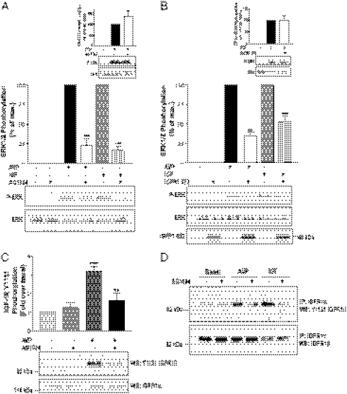
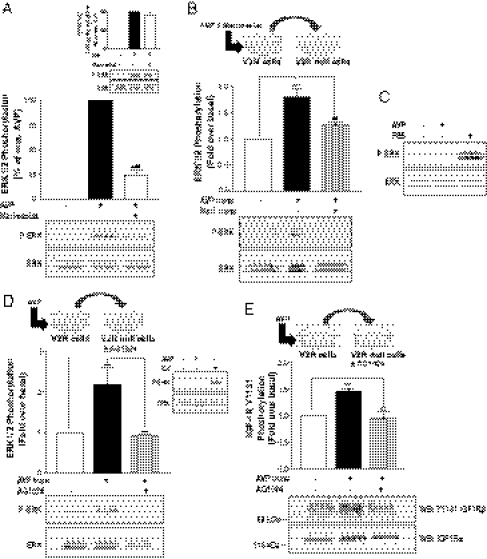
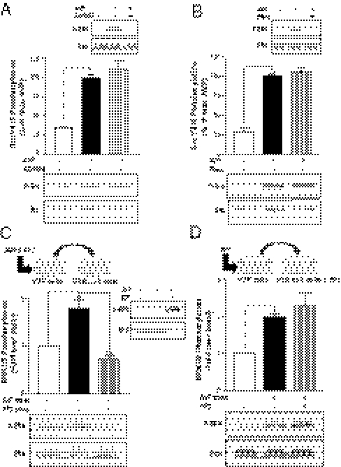
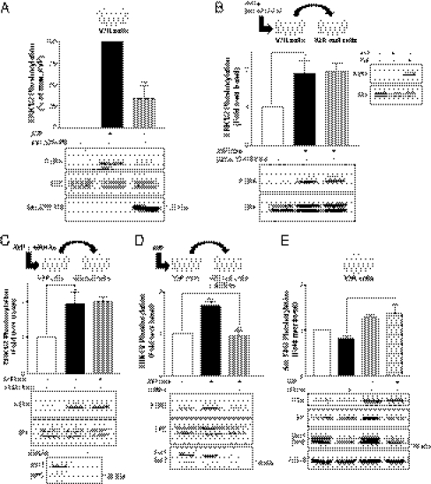
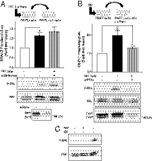
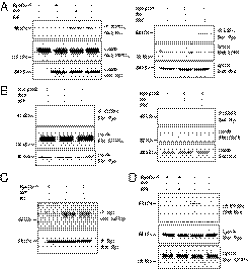
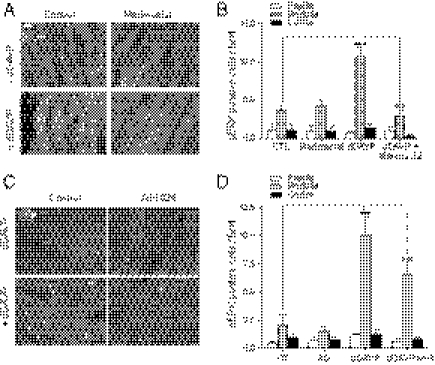
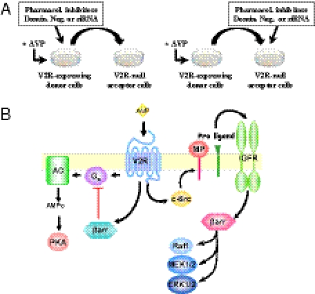
G protein-coupled receptors (GPCRs) have been shown to activate the mitogen-activated protein kinases, ERK1/2, through both G protein-dependent and -independent mechanisms. Here, we describe a G protein-independent mechanism that unravels an unanticipated role for β-arrestins. Stimulation of the V2 vasopressin receptor (V2R) in cultured cells or in vivo in rat kidney medullar collecting ducts led to the activation of ERK1/2 through the metalloproteinase-mediated shedding of a factor activating the insulin-like growth factor receptor (IGFR). This process was found to be both Src- and β-arrestin-dependent. Whereas Src was found to act upstream of the metalloproteinase activation and be required for the release of the IGFR-activating factor, β-arrestins were found to act downstream of the IGFR transactivation. Unexpectedly, the engagement of β-arrestins by the IGFR but not by the V2R was needed to promote the vasopressin-stimulated ERK1/2 activation, indicating that a pool of β-arrestins distinct from those β-arrestins recruited to the V2R acts downstream of the receptor tyrosine kinase to activate ERK1/2. Such a dual site of action for β-arrestins helps explain the pleiotropic actions of this scaffolding protein. Given the role that V2R-stimulated ERK1/2 plays in kidney cell proliferation, this transactivation mechanism may have important implications for renal pathophysiology. Still, the role of β-arrestins downstream of a transactivation event is not limited to the V2R, because we observed a similar involvement for an unrelated GPCR (the platelet-activating factor receptor), indicating that it may be a general mechanism shared among GPCRs.


Conflict of interest statement

The authors declare no conflict of interest.

Figures

Fig. 1.
Fig. 1.
V2R-mediated ERK1/2 activation involves the transactivation of IGFR. (A) Serum-starved HEK293 cells stably expressing V2R were treated or not for 20 min at 37 °C with 5 μM IGFR inhibitor AG1024 before 1 μM AVP or 100 ng/mL IGF1 stimulation for 5 min. MAPK activity was detected by Western blot analysis using phosphospecific anti-ERK1/2 antibody (P-ERK). Expression levels of the MAPK were controlled using antibodies directed against the total kinase population (ERK). Data presented in the bar graph are expressed as a percent of P-ERK/ERK of the level observed in AVP-stimulated conditions. (Inset) EGF-induced ERK1/2 phosphorylation. (B) HEK293 cells transiently expressing myc-V2R were cotransfected or not with the IGFR dominant negative mutant IGFR1-972 and serum-starved before a 1 μM AVP or 100 ng/mL IGF1 stimulation of 5 min. Expression levels of IGFR1-972 were controlled by Western blot using anti-HA antibodies (Lower). (Inset) EGF-induced ERK1/2 phosphorylation. Data represent the mean ± SEM of at least three independent experiments. (C) Serum-starved HEK293 cells stably expressing V2R were pretreated or not for 20 min with 5 μM of the IGFR inhibitor AG1024 and stimulated or not for 5 min at 37 °C with either 1 μM AVP or 100 ng/mL IGF1. Tyrosine autophosphorylation of endogenous IGF1 receptor β-subunit was determined by immunoblotting (WB) whole-cell lysate with anti–IGF-1R pY1131 antibody, and total IGF-1R population was detected with an anti–IGF-1Rα antibody. (D) Endogenous IGFR was immunoprecipitated (IP) from samples derived from cells described in C using the anti–IGF-1Rα 1H7 antibody followed by Western blot (WB) detection of IGF-1R tyrosine autophosphorylation with anti–IGF-1Rβ pY1131 antibody. Total IGF-1R population was detected with an anti–IGF-1Rβ. Typical immunoblots representative of three independent experiments are shown. **P < 0.01; ***P < 0.001.
Fig. 2.
Fig. 2.
V2R-mediated ERK1/2 activation involves a metalloproteinase-dependent transactivation event. (A) Serum-starved HEK293 cells stably expressing V2R were treated or not for 30 min at 37 °C with 10 μM of the metalloproteinase inhibitor marimastat before 1 μM AVP stimulation for 5 min. ERK1/2 phosphorylation was detected and expressed as described in Fig. 1A. (Inset) IGF-induced ERK1/2 phosphorylation. (B) Serum-starved V2R-null cells were incubated for 5 min at 37 °C with the transferred supernatant from V2R-expressing cells previously incubated or not for 30 min with 10 μM marimastat and stimulated or not with AVP (1 μM for 5 min). ERK1/2 phosphorylation was detected as described in Fig. 1A, and data are expressed as fold increase of P-ERK/ERK ratio compared with basal conditions. (C) Serum-starved V2R-null cells were stimulated or not with 1 μM AVP or FBS for 5 min. (D) Serum-starved V2R-null cells previously incubated or not for 20 min with 5 μM AG1024 were incubated for 5 min at 37 °C with the transferred supernatant from V2R-expressing cells stimulated or not with AVP (1 μM for 5 min). ERK1/2 phosphorylation was detected as described in Fig. 1A, and data are expressed as fold increase of P-ERK/ERK ratio compared with basal conditions. (Inset) V2R-null HEK293 cells were stimulated or not with AVP (1 μM) or IGF (100 ng/mL) for 5 min. (E) Serum-starved V2R-null cells previously incubated or not for 20 min with 5 μM AG1024 were incubated for 5 min at 37 °C with the transferred supernatant from V2R-expressing cells stimulated or not with AVP (1 μM for 5 min). Tyrosine autophosphorylation of endogenous IGF1 receptor β-subunit was determined with anti–IGF-1R pY1131 antibody. Total IGF-1R population was detected in cell lysates with the anti–IGF-1Rα antibody. Data represent the mean ± SEM of at least four independent experiments. **P < 0.01; ***P < 0.001.
Fig. 3.
Fig. 3.
V2R-mediated ERK1/2 activation involves an Src-dependent metalloproteinase-mediated transactivation event. (A and B) Serum-starved HEK293 cells stably expressing V2R were pretreated or not with either (A) the IGFR inhibitor AG1024 (5 μM for 20 min) or (B) the metalloproteinase inhibitor 1,10-phenanthroline (500 μM for 30 min) at 37 °C before AVP stimulation (1 μM for 5 min); c-Src phosphorylation at Y416 was detected and quantified using phosphospecific anti-Src (P-Src) and anti-Src GD11 (Src) antibodies. (Insets) AVP-induced ERK1/2 phosphorylation. (C) Serum-starved V2R-null cells were incubated for 5 min at 37 °C with the transferred supernatant from V2R-expressing cells previously incubated or not with PP2 (10 μM for 1 h) and stimulated or not with AVP (1 μM for 5 min). (Inset) V2R-null cells were stimulated or not with AVP or IGF. (D) Serum-starved V2R-null cells pretreated or not with PP2 (10 μM for 1 h) were incubated for 5 min at 37 °C with the transferred supernatant from V2R-expressing cells previously stimulated or not with AVP (1 μM for 5 min). ERK1/2 phosphorylation was detected as described in Fig. 1A, and data are expressed as fold increase of P-ERK/ERK ratio compared with basal conditions. Data represent the mean ± SEM of three independent experiments. *P < 0.05; **P < 0.01; ***P < 0.001.
Fig. 4.
Fig. 4.
β-Arrestins are required downstream of the Src- and metalloproteinase-dependent release of IGFR transactivation factor. (A) HEK293 cells transiently expressing myc-V2R were cotransfected or not with the β-arrestin dominant negative mutant (βarrΔ319–418) and serum-starved before AVP stimulation. Expression levels of βarrΔ319–418 were controlled using the anti–β-arrestin H9 antibody (Lower). ERK1/2 phosphorylation was detected and expressed as described in Fig. 1A. (B) Serum-starved V2R-null cells were incubated for 5 min at 37 °C with the transferred supernatant from HEK293 cells transiently expressing myc-V2R, cotransfected or not with βarrΔ319–418 and previously stimulated or not with AVP (1 μM for 5 min). (Inset) V2R-null cells were stimulated or not with AVP or IGF. (C) Serum-starved V2R-null cells were incubated for 5 min at 37 °C with the transferred supernatant from V2R-expressing cells previously transfected with siRNAs targeting both β-arrestin isoforms or nonspecific siRNA (−) and stimulated or not with AVP (1 μM for 5 min). β-Arrestin knockdown was assessed with the anti–β-arrestin-2 (H9) antibody (Lower). (D) Serum-starved V2R-null cells transfected with siRNAs targeting both β-arrestin isoforms or with nonspecific siRNA (−) were incubated for 5 min at 37 °C with the transferred supernatant from V2R-expressing cells previously stimulated or not with AVP (1 μM for 5 min). ERK1/2 phosphorylation was detected as described in Fig. 1A, and data are expressed as fold increase of P-ERK/ERK ratio compared with basal conditions. (E) HEK293 cells stably expressing V2R were transfected or not with siRNAs targeting both β-arrestin isoforms and serum-starved before AVP stimulation (1 μM for 5 min), and c-Src phosphorylation at Y416 was detected and quantified using phosphospecific anti-Src (P-Src) and anti-Src GD11 (Src) antibodies. β-Arrestin knockdown was assessed with the anti–β-arrestin-1/2 (D24H9) antibody. Data represent the mean ± SEM of at least three independent experiments. *P < 0.05; **P < 0.01; ***P < 0.001.
Fig. 5.
Fig. 5.
β-Arrestins are required downstream of an ectodomain shedding event for PAF-mediated ERK1/2 activation. (A) Serum-starved PAFR-null cells were incubated for 5 min at 37 °C with the transferred supernatant from PAFR-expressing cells previously transfected with siRNAs targeting both β-arrestin isoforms or nonspecific siRNA (−) and stimulated or not with PAF (200 nM for 5 min). (B) Serum-starved PAFR-null cells transfected with siRNAs targeting both β-arrestin isoforms or nonspecific siRNA (−) were incubated for 5 min at 37 °C with the transferred supernatant from PAFR-expressing cells previously stimulated or not with PAF (200 nM for 5 min). β-arrestin knockdown (βarr) was assessed with the anti–β-arrestin-1/2 (D24H9) antibody (A Lower and B Lower). Data represent the mean ± SEM of five independent experiments. (C) PAFR-null cells were stimulated or not with 200 nM PAF or 100 ng/mL IGF for 5 min. ERK1/2 phosphorylation was detected as described in Fig. 1A, and data are expressed as fold increase of P-ERK/ERK ratio compared with basal conditions. *P < 0.05; **P < 0.01.
Fig. 6.
Fig. 6.
V2R-promoted association of β-arrestins to the transactivated IGFR. (A and B) HEK293 cells stably expressing V2R were transiently transfected or not with either myc-β-arrestin-1 (A) or myc-β-arrestin-2 (B) and serum-starved before a 5-min AVP (1 μM), IGF (100 ng/mL), or EGF (10 ng/mL) stimulation at 37 °C. Cells were then subjected to immunoprecipitation (IP) with an antibody specific to the α-subunit of the IGF-1R (Left) or an antibody specific to the N terminus of EGFR (Right). Coimmunoprecipitation of β-arrestins was assessed by blotting with an anti-myc antibody. Endogenous IGFR and EGFR expression levels were detected in cell lysates with the anti–IGF-1Rα and the anti-EGFR antibodies, respectively. Expression levels of the transfected constructs were detected in cell lysates using an anti-myc antibody. (C) myc-β-arrestin-1 was immunoprecipitated using an anti-myc antibody, and the presence of IGFR and β-arrestin-1 were probed using anti-IGFRβ and anti-myc antibodies, respectively. (D) HEK293 cells stably expressing V2R were transiently transfected with myc-β-arrestin-1, serum-starved, and pretreated or not with PP2 (10 μM for 1 h) before AVP (1 μM for 5 min) stimulation at 37 °C. Endogenous IGF-1R was then immunoprecipitated, and β-arrestin-1 association was detected with anti-Myc antibody. Typical immunoblots representative of at least three independent experiments are shown.
Fig. 7.
Fig. 7.
The phosphorylation-deficient V2R(Ala6) conserves its ability to stimulate ERK1/2 activity in a β-arrestin–dependent manner. (A) Serum-starved HEK293 cells stably expressing either WT-V2R or V2R(Ala6) were stimulated at 37 °C with 1 μM AVP for the indicated times. (B) HEK293 cells stably expressing V2R(Ala6) were transfected with siRNAs targeting both β-arrestin isoforms (dotted line) or not (solid line) and serum-starved before 1 μM AVP stimulation at 37 °C for the indicated times. ERK1/2 phosphorylation was detected and expressed as described in Fig. 1A. (Inset) β-arrestin knockdown was assessed with the anti–β-arrestin-2 (H9) antibody.
Fig. 8.
Fig. 8.
The AVP-stimulated ERK1/2 activity in the rat kidney is metalloproteinase- and IGFR-dependent. Phospho-ERK1/2 activity was assessed in kidney slices from rat treated or not (control) for 3 d with dDAVP (0.13 μg/rat per day) in the absence or presence of (A) marimastat (3.6 mg/rat per day) or (C) AG1024 (0.30 mg/rat per day) using an antiphospho-ERK1/2 antibody. pERK activity was detected in the rat kidney medulla by epifluorescence microscopy (objective = 40×). (B and D) The histograms depict the number of pERK1/2-positive cells detected per confocal microscope field (20× objective) in 50-μm-thick slices of rat papilla (white), medulla (gray), or cortex (black) after the indicated treatment. Three to four kidney sections were evaluated for each animal, and three animals were tested for each condition. The data shown represent the mean ± SEM of three independent experiments. ***P < 0.001.
Fig. P1.
Fig. P1.
(A) Supernatant transfer assay. The supernatant transfer assay used to dissect the molecular events upstream and downstream of IGFR transactivation. V2R-null acceptor cells were stimulated with the transferred supernatant of AVP-stimulated, V2R-expressing donor cells that were or were not pretreated with inhibitors, dominant negative mutants, or siRNAs. (B) V2R-regulated signaling. V2R-regulated β-arrestin–dependent activities where AVP binding to V2R elicits the engagement of distinct β-arrestin (βarr) pools involved in opposed signaling events. AVP stimulation of V2R induces the Src-dependent metalloproteinase-mediated (MP) shedding of an IGFR transactivating factor, promoting β-arrestin recruitment to IGFR and the ensuing ERK1/2 activity. However, recruitment of β-arrestins to the activated V2R leads to signal arrest through desensitization of the Gs-adenylate cyclase (AC) –PKA signaling pathway.

References

    1. Charest PG, Bouvier M. Palmitoylation of the V2 vasopressin receptor carboxyl tail enhances beta-arrestin recruitment leading to efficient receptor endocytosis and ERK1/2 activation. J Biol Chem. 2003;278:41541–41551. - PubMed
    1. Tohgo A, et al. The stability of the G protein-coupled receptor-beta-arrestin interaction determines the mechanism and functional consequence of ERK activation. J Biol Chem. 2003;278:6258–6267. - PubMed
    1. Charest PG, Oligny-Longpré G, Bonin H, Azzi M, Bouvier M. The V2 vasopressin receptor stimulates ERK1/2 activity independently of heterotrimeric G protein signalling. Cell Signal. 2007;19:32–41. - PubMed
    1. Werry TD, Sexton PM, Christopoulos A. “Ins and outs” of seven-transmembrane receptor signalling to ERK. Trends Endocrinol Metab. 2005;16:26–33. - PubMed
    1. Wetzker R, Böhmer FD. Transactivation joins multiple tracks to the ERK/MAPK cascade. Nat Rev Mol Cell Biol. 2003;4:651–657. - PubMed

Publication types

LinkOut - more resources