. 2012 May 1;109(18):E1072-81.
doi: 10.1073/pnas.1120320109. Epub 2012 Apr 9.

Drosophila Golgi membrane protein Ema promotes autophagosomal growth and function

Affiliations

Drosophila Golgi membrane protein Ema promotes autophagosomal growth and function

Sungsu Kim et al. Proc Natl Acad Sci U S A. .

Abstract

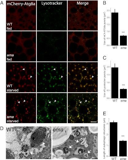
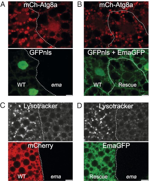
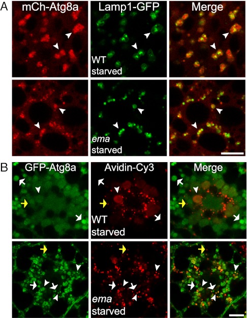
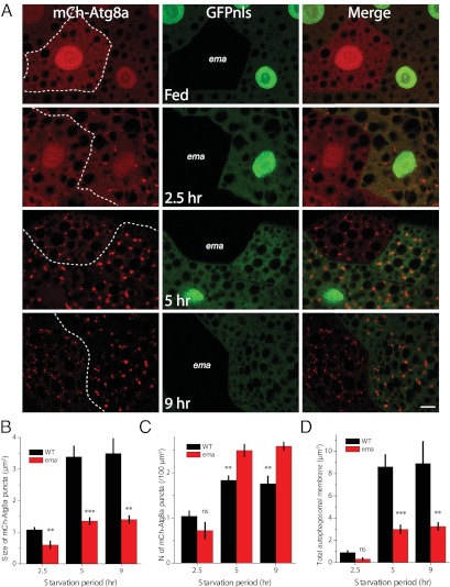
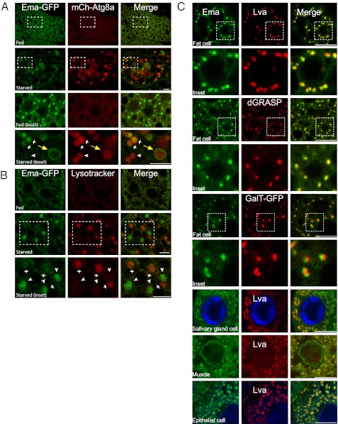
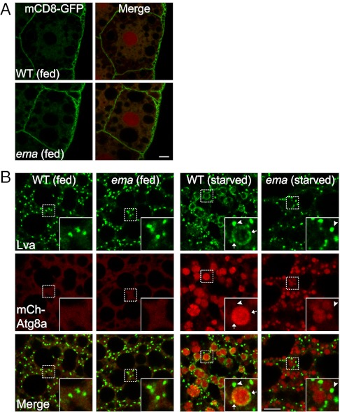
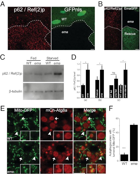
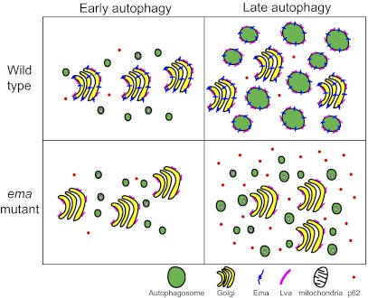
Autophagy is a self-degradative process in which cellular material is enclosed within autophagosomes and trafficked to lysosomes for degradation. Autophagosomal biogenesis is well described; however mechanisms controlling the growth and ultimate size of autophagosomes are unclear. Here we demonstrate that the Drosophila membrane protein Ema is required for the growth of autophagosomes. In an ema mutant, autophagosomes form in response to starvation and developmental cues, and these autophagosomes can mature into autolysosomes; however the autophagosomes are very small, and autophagy is impaired. In fat body cells, Ema localizes to the Golgi complex and is recruited to the membrane of autophagosomes in response to starvation. The Drosophila Golgi protein Lva also is recruited to the periphery of autophagosomes in response to starvation, and this recruitment requires ema. Therefore, we propose that Golgi is a membrane source for autophagosomal growth and that Ema facilitates this process. Clec16A, the human ortholog of Ema, is a candidate autoimmune susceptibility locus. Expression of Clec16A can rescue the autophagosome size defect in the ema mutant, suggesting that regulation of autophagosome morphogenesis may be a fundamental function of this gene family.


Conflict of interest statement

The authors declare no conflict of interest.

Figures

Fig. 1.
Fig. 1.
Ema is required for formation of normal autophagosomes. (A) ema-mutant autophagosomes are smaller than wild type but can mature into acidic autolysosomes. Shown are representative single confocal sections of Drosophila fat body cells expressing the autophagosomal marker mCherry-Atg8a (red) and stained with LysoTracker DND-26 (green) from early third-instar larvae fed on yeast extract-enriched fly food or starved in EBSS buffer for 6 h. Arrowheads indicate LysotTracker-positive autophagosomes. Wild type = CG-Gal4/+,UAS-mCherry-Atg8a/+; ema = CG-Gal4/+,UAS-mCherry-Atg8a/+,ema1. (Scale bar, 10 μm.) (B and C) Quantification of the size of autophagic structures in A: mCherry-Atg8a autophagosomes (B) and LysoTracker-positive autolysosomes (C). (D and E) Ultrastructural analysis confirms the presence of small autophagosomes in the ema mutant. (D) Representative electron micrographs of starved Drosophila fat body cells. Autophagic structures (arrows) contain remnants of rough ER and mitochondria. (Scale bars, 2 μm.) (E) Quantification of the length of autophagosomes in D. n = 219 autophagic structures from eight sections from wild type; n = 326 from 12 sections from ema mutant. Data in B, C, and E represent mean ± SE; ***P < 0.001 (t test).
Fig. 2.
Fig. 2.
Ema acts cell autonomously for autophagy. (A and B) Cell-autonomous function of ema for normal autophagosome formation. (A) Representative single confocal section of starved mosaic fat body cell clones expressing the autophagosomal marker mCherry-Atg8a (red). Expression of nuclear GFP (GFPnls, green) marks wild-type clones. Genotype of the animal: hs-flp/+;CG-Gal4/+;UAS-mCherry-Atg8a, FRT82B,UAS-GFPnls/FRT82B,ema1. (B) Representative single confocal section of starved mosaic fat body cell clones expressing the autophagosomal marker mCherry-Atg8a (red) and Ema-GFP (green). Note expression of GFPnls (green) in the wild-type clone but not in the ema-mutant clone. Genotype of the animal: hs-flp/+;CG-Gal4/+;UAS-Ema-GFP/+;UAS-mCherry-Atg8a, FRT82B,UAS-GFPnls/FRT82B,ema1. (C and D) Cell-autonomous function of ema for normal autolysosome formation. (C) Representative live single confocal section of starved mosaic fat body cell clones stained with LysoTracker DND-26. mCherry expression marks wild-type clones (red). Genotype of the animal: hs-flp/+;r4-Gal4,FRT82B,UAS-mCherry/FRT82B,ema1. (D) Representative live single confocal sections of starved mosaic fat body cell clones stained with LysoTracker DND-99. Ema-GFP (green) marks rescue clone. Genotype of the animal: da-Gal4,UAS-Ema-GFP, ema1. (Scale bars, 10 μm.)
Fig. 3.
Fig. 3.
ema is dispensable for autophagosomal fusion with endosomes and lysosomes. (A) Autophagosomal fusion with lysosomes. Representative single confocal sections show part of a starved Drosophila fat body cell that expresses both the lysosomal marker Lamp1-GFP (green) and the autophagosomal marker mCherry-Atg8a (red). Arrowheads indicate autolysosomes (fusion products of autophagosome and lysosome) that contain both Lamp1-GFP and mCherry-Atg8a. Wild type = CG-Gal4/+,UAS-Lamp1-GFP/+,UAS-mCherry-Atg8a/+; Ema = CG-Gal4/+,UAS-Lamp1-GFP/+,UAS-mCherry-Atg8a/+, ema1. (B) Autophagosomal fusion with endosomes. Representative single confocal sections of fat body cells expressing the autophagosomal marker mCherry-Atg8a (green) and labeled with the endocytic tracer avidin-Cy3 (red). Arrowheads indicate an amphisome (fusion product of autophagosome and endosome) that contains both GFP-Atg8a and avidin-Cy3. White arrows indicate autophagosomes that have not fused with endosomes. Yellow arrows indicate an endosome that has not fused with autophagosomes. Wild type = da-Gal4/+,UAS-GFP-Atg8a/+; Ema = da-Gal4/+,UAS-GFP-Atg8a/+,ema1.
Fig. 4.
Fig. 4.
Ema promotes autophagosomal growth. Small autophagosomes in the ema mutant result from defective autophagosomal growth. (A) Representative single confocal sections of mosaic fat body cell clones expressing the autophagosomal marker mCherry-Atg8a (red) at the indicated starvation periods. Expression of GFPnls (green) marks wild-type clones. Genotype of the animal: hs-flp/+;CG-Gal4/+;UAS-mCherry-Atg8a, FRT82B,UAS-GFPnls/FRT82B,ema1. (Scale bar, 10 μm.) (B and C) Quantification of the size (B) and number (C) of autophagosomes. (D) Total membrane (spherical volume; 4/3Πr3) of autophagosomes in A (n = 7 animals at 2.5 h, 8 animals at 5 h, and 5 animals at 9 h). Data represent mean ± SE (**P < 0.01; ***P < 0.001, ANOVA). ns, not significant.
Fig. 5.
Fig. 5.
Ema localizes to the Golgi complex and moves to autophagosomes under starvation conditions. (A and B) Localization of Ema protein to autophagosomes during autophagy. (A) Representative single confocal sections of fed and starved fat body cells expressing Ema-GFP (green) and mCherry-Atg8a (red). Insets show autophagosomes with (white arrows) or without (arrowhead) Ema-GFP. Note that Ema-GFP localizes at the perimeter of autophagosomes (yellow arrows). (B) Representative single confocal sections of fed and starved fat body cells expressing Ema-GFP (green) and stained with LysoTracker DND-99 (red). Insets show the lack of colocalization of Ema-GFP (arrowheads) and LysoTracker structures (arrows). (C) Ema protein localizes to the Golgi complex. Representative single confocal sections of fat body cells, salivary gland cells, muscles, and epithelial cells expressing Ema protein tagged with either GFP (green) or mCherry(red) and labeled for the Golgi proteins Lva, dGRASP-GFP, or GalT-GFP. (Scale bars, 10 μm.)
Fig. 6.
Fig. 6.
Defective Golgi-to-autophagosome traffic in the ema fat body cell. (A) ema does not affect general Golgi membrane trafficking. Shown are representative single confocal sections of fat body cells expressing the plasma membrane protein mCD8-GFP. Note the proper localization of mCD8-GFP at the cell surface in both wild-type and ema-mutant cells. mCherry-Atg8a (red) was used as reference for the cell nuclei. (B) Recruitment of the Golgi protein Lva to autophagosomes requires ema. Shown are representative single confocal sections of fat body cells expressing mCherry-Atg8a (red) and labeled for Lva protein (green). Insets show large and small Lva structures (indicated by arrowheads and arrows, respectively) separated from or associated with mCherry-Atg8a autophagosomes of starved cells. (Scale bars, 10 μm.)
Fig. 7.
Fig. 7.
Ema is required for autophagy of p62 and mitochondria. (AD) Ema promotes p62 turnover. (A) Representative confocal images of starved fat body cell clones labeled for p62 protein (red). GFPnls (green) marks wild-type clones. Genotype of the animal: hs-flp/+;r4-Gal4,FRT82B,UAS-mCherry/FRT82B,ema1. n = 10 pairs of mosaic clones. *P < 0.05 (paired t test). (B) Representative confocal images of starved fat body cell clones labeled for p62 protein (red). Ema-GFP (green) marks rescue clones. Genotype of the animal: da-Gal4,UAS-Ema-GFP, ema1. n = 7 pairs of mosaic clones. (C) Immunoblot of p62 protein in whole-animal lysates. The p62 level is normalized to β-tubulin. n = 5 independent Western blot experiments. (D) Quantifications of p62 level in A, B, and C (mean ± SE). *P < 0.05, (paired t test). ns, not significant. (E and F) Ema promotes mitophagy. (E) Representative single confocal sections of starved fat body cells expressing the mitochondria marker Mito-GFP (green) and autophagosomal marker mCherry-Atg8a (red). Arrowheads indicate small autophagosomes; arrows indicate luminal Mito-GFP puncta in the large autophagosomes. Left Inset depicts small autophagosomes in both wild-type and mutant fat body cells containing Mito-GFP puncta (note very strong luminal Mito-GFP fluorescence in the ema mutant). Right Inset depicts luminal Mito-GFP puncta in the large autophagosomes in the mutant but not in the wild type. (F) Quantification of the fraction (%) of autophagosomes with luminal Mito-GFP puncta in E (mean ± SE). ***P < 0.001 (t test). (Scale bars, 10 μm.)
Fig. P1.
Fig. P1.
Schematic diagram of autophagosomal growth in Drosophila fat body cells. In wild-type fat body cells, small autophagosomes form upon autophagy induction and grow into large autophagosomes. The transmembrane protein Ema and the peripheral membrane-associated protein Lva reside on the Golgi complex in the steady state and move onto these growing autophagosomes. In the ema-mutant cells, small autophagosomes form upon autophagy induction but fail to grow, and Lva is not recruited to autophagosomes. Autophagic turnover of the p62 protein and of mitochondria is impaired in the ema mutant. Thus, ema is required for both autophagosomal growth and autophagic function. These findings are consistent with a model in which ema promotes autophagosomal growth by promoting Golgi-to-autophagosomal membrane traffic.

References

    1. He C, Klionsky DJ. Regulation mechanisms and signaling pathways of autophagy. Annu Rev Genet. 2009;43:67–93. - PMC - PubMed
    1. Berry DL, Baehrecke EH. Growth arrest and autophagy are required for salivary gland cell degradation in Drosophila. Cell. 2007;131:1137–1148. - PMC - PubMed
    1. Mizushima N, Levine B, Cuervo AM, Klionsky DJ. Autophagy fights disease through cellular self-digestion. Nature. 2008;451:1069–1075. - PMC - PubMed
    1. Levine B, Mizushima N, Virgin HW. Autophagy in immunity and inflammation. Nature. 2011;469:323–335. - PMC - PubMed
    1. Suzuki K, Ohsumi Y. Current knowledge of the pre-autophagosomal structure (PAS) FEBS Lett. 2010;584:1280–1286. - PubMed

Publication types

MeSH terms