. 2012 Apr 24;109(17):6704-9.
doi: 10.1073/pnas.1118058109. Epub 2012 Apr 9.

Anticancer drug oxaliplatin induces acute cooling-aggravated neuropathy via sodium channel subtype Na(V)1.6-resurgent and persistent current

Affiliations

Anticancer drug oxaliplatin induces acute cooling-aggravated neuropathy via sodium channel subtype Na(V)1.6-resurgent and persistent current

Ruth Sittl et al. Proc Natl Acad Sci U S A. .

Abstract

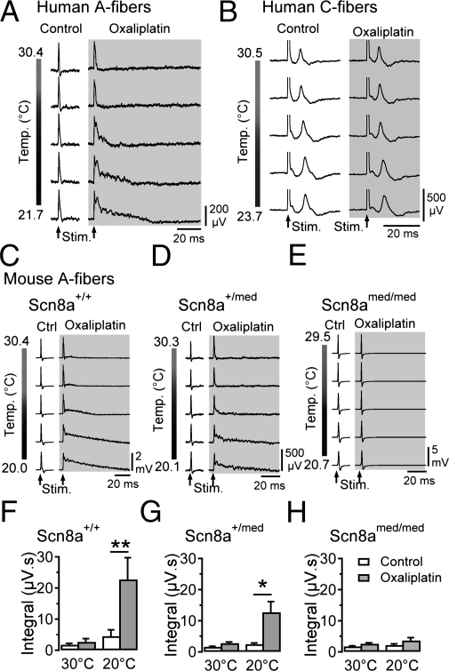
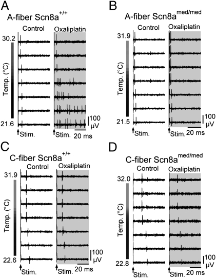
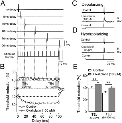
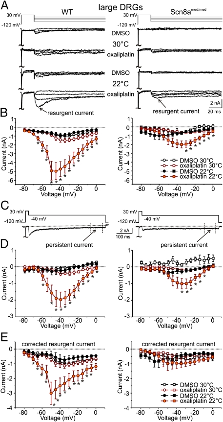
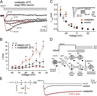
Infusion of the chemotherapeutic agent oxaliplatin leads to an acute and a chronic form of peripheral neuropathy. Acute oxaliplatin neuropathy is characterized by sensory paresthesias and muscle cramps that are notably exacerbated by cooling. Painful dysesthesias are rarely reported for acute oxaliplatin neuropathy, whereas a common symptom of chronic oxaliplatin neuropathy is pain. Here we examine the role of the sodium channel isoform Na(V)1.6 in mediating the symptoms of acute oxaliplatin neuropathy. Compound and single-action potential recordings from human and mouse peripheral axons showed that cooling in the presence of oxaliplatin (30-100 μM; 90 min) induced bursts of action potentials in myelinated A, but not unmyelinated C-fibers. Whole-cell patch-clamp recordings from dissociated dorsal root ganglion (DRG) neurons revealed enhanced tetrodotoxin-sensitive resurgent and persistent current amplitudes in large, but not small, diameter DRG neurons when cooled (22 °C) in the presence of oxaliplatin. In DRG neurons and peripheral myelinated axons from Scn8a(med/med) mice, which lack functional Na(V)1.6, no effect of oxaliplatin and cooling was observed. Oxaliplatin significantly slows the rate of fast inactivation at negative potentials in heterologously expressed mNa(V)1.6r in ND7 cells, an effect consistent with prolonged Na(V) open times and increased resurgent and persistent current in native DRG neurons. This finding suggests that Na(V)1.6 plays a central role in mediating acute cooling-exacerbated symptoms following oxaliplatin, and that enhanced resurgent and persistent sodium currents may provide a general mechanistic basis for cold-aggravated symptoms of neuropathy.


Conflict of interest statement

The authors declare no conflict of interest.

Figures

Fig. 1.
Fig. 1.
Oxaliplatin and cooling-induced CAP-afterpotentials require NaV1.6. Exposure of segments of human (A and B) and mouse (CE) sural nerve to oxaliplatin (100 μM; ∼90 min; gray) induces the emergence of after-potentials upon cooling following electrically evoked (Stim.) CAP responses in A-fibers (A, C, and D, n = 8 for human, and n = 55 for mouse nerve segments). The C-fiber CAP for a human nerve fascicle (B) that fortuitously contained very few functional A-fibers remains unaffected during cooling both before and after exposure to oxaliplatin. Cooling following oxaliplatin (gray) alters the shape of A-fiber responses from wild-type mice (Scn8a+/+; C, n = 8 at 30 °C, 7 at 20 °C) and heterozygous (Scn8a+/med; D, n = 21 at 30 °C, 16 at 20 °C), but not Scn8amed/med mice lacking NaV1.6 (E, n = 21 at 30 °C, 16 at 20 °C). The magnitude of after-potential activity, quantified by integrating the voltage response following electrical stimulation (μV.s), is larger in recordings from wild-type mice than in those from Scn8a+/med mice (F–H). (*P < 0.05).
Fig. 2.
Fig. 2.
Axonal effects of oxaliplatin and cooling are A-fiber specific and require NaV1.6. Responses of individual A- and C-fibers from wild-type (Scn8a+/+) and Scn8amed/med mice during cooling under control conditions and following exposure to oxaliplatin (100 μM; ∼90 min; gray) show that cooling results in the induction of stimulus-evoked repetitive AP discharge selectively in single myelinated (A) but not unmyelinated (C) axons in wild-type mice. Single myelinated (B) and unmyelinated (D) axons from Scn8amed/med mice, lacking functional NaV1.6, show no stimulus-evoked repetitive discharge upon cooling, highlighting the importance of Nav1.6 for cooling-induced burst discharge.
Fig. 3.
Fig. 3.
Enhancement of depolarizing threshold electrotonus (TE) responses by oxaliplatin is positively correlated with repetitive after-activity in A-fibers. Changes in electrical threshold in response to polarizing current (TE) applied to myelinated axons. (A) The current amplitude required to evoke a 40% A-fiber CAP under control conditions (black uppermost trace and black vertical line in A, black marker in B) is used as a reference (horizontal broken black line in stimulus current trace). The current strength required to similarly evoke a 40% CAP response at discrete time points (delays) during application of polarizing current (A, stimulus current) was determined in sequential sweeps (gray traces). In the example illustrated in A, a conditioning depolarizing current of +40% of the control threshold current is applied over 100 ms. Individual sweeps show 40% CAP responses evoked at the indicated delay. (B) Representative examples of TE responses to ±40% polarizing current before (Control open markers) and after oxaliplatin (100 μM, ∼90 min; filled gray markers) with the periods used for quantitative analysis is indicated in E (d, depolarizing; h, hyperpolarizing). (C and D) Representative examples of CAP responses to electrical stimulation during conditioning subthreshold depolarizing (C) and hyperpolarizing (D) current application (Lowermost trace) in the absence (Control, Upper trace) and presence of oxaliplatin (Middle traces, gray). (E) Pooled data for +40% depolarizing TE responses determined over 10–20 ms and 90–100 ms in the presence of vehicle (control) and oxaliplatin. Data were collected at a bath temperature of 25.5 ± 1 °C from nine wild-type mice aged 122–194 d and weighing between 24.4 and 34.0 g. (**P < 0.01).
Fig. 4.
Fig. 4.
Oxaliplatin and cooling enhance INaR and INaP TTX-s Nav currents in large-diameter DRG neurons. (A) Representative current traces in response to voltage commands to −75, −45, −25, and −5 mV (Upper lane) from large-diameter (40.6 ± 4.5 μm, n = 25) DRG neurons from wild-type (WT, Left) and Scn8amed/med mice (Right) at 30 °C and 22 °C after incubation with oxaliplatin (30 μM; ∼90 min). (B) Peak INaR as a function of voltage for recordings at 30 °C [open symbols: WT n = 5;5 and Scn8amed/med: n = 4;6 (DMSO; oxaliplatin)] and 22 °C (filled symbols, WT: n = 10;11, Scn8amed/med: n = 6;8) from DRG neurons incubated with vehicle (1% vol/vol DMSO) or oxaliplatin. (C) Representative voltage command (Upper) and current (Lower) traces indicating the period over which the INaP component was determined (400–475 ms). (D) INaP shown as a function of voltage for recordings at 30 °C (open symbols, WT n = 5;5 and Scn8amed/med n = 8;11 for DMSO; oxaliplatin) and 22 °C (filled symbols, WT: n = 10;11, Scn8amed/med: n = 6;8) from large-diameter DRG neurons (WT Left, Scn8amed/med Right) after incubation with vehicle (black) or oxaliplatin (orange). (E) Corrected INaR amplitude as a function of voltage for recordings at 30 °C (open) and 22 °C (filled) from large-diameter DRG neurons (WT Left, Scn8amed/med Right) after incubation with vehicle (black) or oxaliplatin (orange). INaR was corrected for each individual trace by subtracting the INaP component (D) from the peak INaR (B). Mice ranging in age from P14–25 were used. (*P < 0.05, **P < 0.01).
Fig. 5.
Fig. 5.
Oxaliplatin slows the rate of decay of resurgent current in large DRG neurons and slows fast inactivation in mNaV1.6r in ND7 cells. (A) Representative current recordings (Lower) from a DRG neuron in response to the voltage protocol (Upper). Current traces were fitted with a double-exponential function. (B) Rise (τ1, triangles) and decay (τ2, circles) time constants for resurgent current evoked by the protocol in A as a function of voltage at 22 °C (DMSO: black symbols, n = 7; oxaliplatin 30 μM: orange symbols, n = 10). (C) Time constant of mNaV1.6r current inactivation as a function of voltage. mNaV1.6r was expressed with β4 in ND7 cells and currents were evoked by voltage steps from −90 mV to potentials ranging from −60 to +30 mV (control: black symbols, n = 29; oxaliplatin 30 μM: orange symbols, n = 28). (Inset) Depiction of voltage commands (Lower Right) and current recordings. All recordings were performed at room temperature (∼22 °C). (D) Simplified NaV gating schematic indicating state transitions during the voltage protocol used to evoke resurgent current in DRG neurons. At −120 mV, NaVs are closed (dark gray) and open (white) upon depolarization to +30 mV. At positive potentials, open channels may either inactivate (arrow 1, Upper) or, if an appropriate blocking particle (black circle) is present, block in the open configuration (Lower). Upon partial repolarization to voltages between 0 and −60 mV, the blocking particle is expelled by permeating Na+ generating a resurgent current (INaR). Resurgent current is terminated either by fast inactivation (arrow 2) or deactivation (not shown). In mNav1.6r, oxaliplatin slowed fast inactivation voltage dependently at potentials in the range of INaR (C). (E) Comparison of DRG current recordings (gray) and mathematical simulations (black and red). To mimic the empirical slowing of fast inactivation by oxaliplatin (Lower gray trace), the rate constant (Oon) for the state transition from open (O) to inactivated (I6) was reduced to 2% of its original value (0.02 × Oon), resulting in an increase in the magnitude of simulated resurgent current and the emergence of a persistent current component. (*P < 0.05, **P < 0.01).

References

    1. Lehky TJ, Leonard GD, Wilson RH, Grem JL, Floeter MK. Oxaliplatin-induced neurotoxicity: Acute hyperexcitability and chronic neuropathy. Muscle Nerve. 2004;29:387–392. - PubMed
    1. Hill A, et al. Detecting acute neurotoxicity during platinum chemotherapy by neurophysiological assessment of motor nerve hyperexcitability. BMC Cancer. 2010;10:451. - PMC - PubMed
    1. Raymond E, Chaney SG, Taamma A, Cvitkovic E. Oxaliplatin: A review of preclinical and clinical studies. Ann Oncol. 1998;9:1053–1071. - PubMed
    1. Gauchan P, Andoh T, Kato A, Kuraishi Y. Involvement of increased expression of transient receptor potential melastatin 8 in oxaliplatin-induced cold allodynia in mice. Neurosci Lett. 2009;458:93–95. - PubMed
    1. Nassini R, et al. Oxaliplatin elicits mechanical and cold allodynia in rodents via TRPA1 receptor stimulation. Pain. 2011;152:1621–1631. - PubMed

Publication types

LinkOut - more resources