. 2012 Apr 24;109(17):6626-31.
doi: 10.1073/pnas.1204520109. Epub 2012 Apr 9.

Using translational enhancers to increase transgene expression in Drosophila

Affiliations

Using translational enhancers to increase transgene expression in Drosophila

Barret D Pfeiffer et al. Proc Natl Acad Sci U S A. .

Abstract

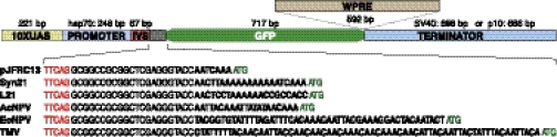
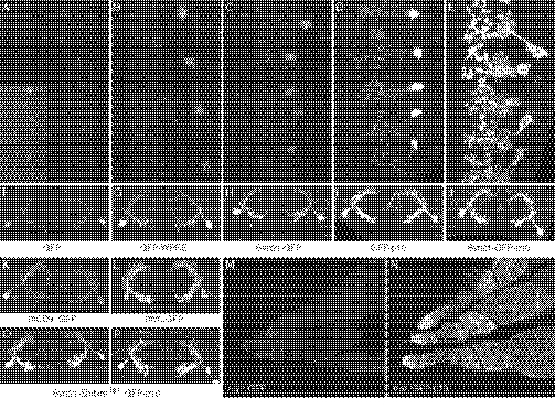
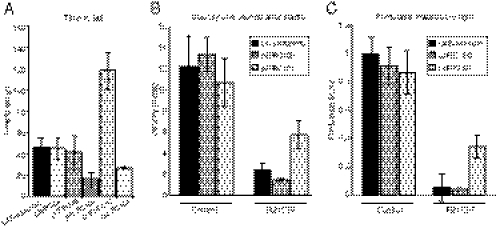
The ability to specify the expression levels of exogenous genes inserted in the genomes of transgenic animals is critical for the success of a wide variety of experimental manipulations. Protein production can be regulated at the level of transcription, mRNA transport, mRNA half-life, or translation efficiency. In this report, we show that several well-characterized sequence elements derived from plant and insect viruses are able to function in Drosophila to increase the apparent translational efficiency of mRNAs by as much as 20-fold. These increases render expression levels sufficient for genetic constructs previously requiring multiple copies to be effective in single copy, including constructs expressing the temperature-sensitive inactivator of neuronal function Shibire(ts1), and for the use of cytoplasmic GFP to image the fine processes of neurons.


Conflict of interest statement

The authors declare no conflict of interest.

Figures

Fig. 1.
Fig. 1.
Structures of UAS-GFP constructs used in this study. Most components have been held constant and correspond to those in the previously described pJFRC13 vector (29), including the binding sites for the transactivator GAL4 (10XUAS), the promoter (a fragment of the hsp70 gene), a small intron (IVS; derived from the Drosophila myosin heavy chain gene), a GFP coding region optimized for Drosophila codon use, and the transcription terminator sequence derived from SV40. The sizes of these components are indicated. In the constructs used to assay the effects of 5′-UTR elements on GFP levels (see Fig. 2), the nucleotides immediately upstream of the ATG initiating codon of GFP in pJFRC13 have been replaced. The nucleotide sequences of this region, which spans from the end of the IVS (TTCAG, shown in red) to the initiating ATG (shown in green), are shown for each of the constructs. Only the nucleotides shown in bold differ between constructs: pJFRC13 (29); Syn21, pJFRC80 (see Methods); L21, pJFRC83 (35); AcNPV, pJFRC84 (10); EoNPV, pJFRC85 (9); and TMV, pJFRC86 (11). To make the constructs used to test the effects of altering the 3′-UTR, either the WPRE element (33) was placed between the GFP gene and the SV40 terminator (GFP-WPRE) or the SV40 terminator was replaced by the terminator from the AcNPV p10 gene (GFP-p10) (25). The constructs were all inserted in the same genomic location (attP2) to equalize extrinsic effects on expression.
Fig. 2.
Fig. 2.
Short 5′-UTR sequence elements derived from a variety of viral and cellular mRNAs can increase protein expression in Drosophila. Ventral nerve cords of third-instar larvae that each carry the R66A12-GAL4 driver, paired with a different 10XUAS-GFP construct, are shown; the preparations have been stained with an antibody against GFP. R66A12 drives expression in a sparse set of segmentally repeated neurons, allowing the cell bodies (located laterally) and neurites (located centrally) of individual neurons to be resolved. (A) Expression level obtained with pJFRC13 (29). (BF) Increased expression of GFP obtained by including in the 5′-UTR the additional sequence: Syn21 (B); L21 (C); AcNPV (D); EoNPV (E); and TMV (F). See Fig. 1 for a more detailed description of the constructs.
Fig. 3.
Fig. 3.
Enhancement of protein expression levels by 3′-UTR elements. (AE) Increase in the level of cytoplasmic GFP generated from 10XUAS constructs by the addition of various 5′- or 3′-UTR elements was determined by measuring native GFP fluorescence using the photomultiplier tube (PMT) of a Zeiss 510 confocal microscope. Dissected nervous systems were imaged under identical conditions except for the Inset in A, which was imaged at higher gain. (A) Level of expression obtained with pJFRC13 (Fig. 1) (29), which was used as a baseline to measure the level of enhancement produce by addition of the other sequence elements. (B) Increase of 6.1-fold in cell body GFP fluorescence was observed when the WPRE element was added to the 3′-UTR in pJFRC14 (29). (C) Increase of 7.5-fold was observed by the addition of the Syn21 element to the 5′-UTR (pJFRC80). (D) p10 element produces a 23-fold increase when added to the 3′-UTR (pJFRC28). (E) Further increases in expression are observed when both the Syn21 and p10 elements are present (pJFRC81). (FJ) Pairs of neurons, viewed transversely, from the same series of genotypes, following staining with anti-GFP antibody. (K and L) Specimens imaged as in FJ using vectors that contain the same UTR elements as pJFRC13 but that express membrane-targeted GFP, either mCD8::GFP (K; pJFRC2; 29) or myr::GFP (L; pJFRC12; 29). M (pJFRC12) and N (pJFRC29) show that the p10 element can also increase GFP expression in the female germline. (O and P) Antibody staining for GFP in lines carrying a 20XUAS-Syn21-Shibire[ts1]::GFP-p10 construct (pJFRC101) inserted at the VK00005 (O) or attP2 (P) genomic integration sites. The R66A12-GAL4 driver was used to generate the data shown in AL and O and P. The R34C10-GAL4 driver was used for M and N.
Fig. 4.
Fig. 4.
Assays of the effectiveness of Shibirets1 constructs. (A) Ability of various pJFRC constructs (in VK00005) that express Shibirets1 to induce loss of motor control was compared with that of the Kitamoto stock. pJFRC98, pJFRC99, pJFRC100, and pJFRC101 were crossed to a pan-neuronal GAL4 driver, R57C10-GAL4, and pJFRC104 was crossed to R57C10-LexAp65 (in VK00027). The times for flies to fall from the side of the vial at 30.6 °C are shown (mean ± SD). The progeny of control crosses of the same effectors to the BDPGal4U line, which does not express significant amounts of GAL4 in the adult nervous system (36), were all still on the side of the vial after 360 seconds, when the experiment was terminated. The pJFRC effector constructs were inserted into the VK00005 genomic site; see Table 1 for details of the constructs. (B) Effectiveness of the pJFRC100, pJFRC101, and Kitamoto stocks in decreasing locomotor response to startle was compared (all values are mean ± SD). The effectors were each crossed to the control GAL4 driver, BDPGal4U, or to a driver line, R21C07-GAL4, that was expressed in the optic lobes and a small proportion of central brain and ventral nerve cord neurons. Flies normally respond to a startle by an increase in locomotion. The extent to which each of the effector lines was able to suppress this response is shown. (C) Flies show a strong phototaxis response to UV light. The same genotypes as in B were assayed for phototaxis. A phototaxis score was calculated by averaging the median displacement of six tubes of flies toward lights and normalizing by the length of the tubes (mean ± SD). This is a normalized metric for the strength of attraction toward light where a value of one indicates that the population of flies congregates as close to the light as possible and a value of zero indicates no attraction.

References

    1. Gray NK, Wickens M. Control of translation initiation in animals. Annu Rev Cell Dev Biol. 1998;14:399–458. - PubMed
    1. Jackson RJ, Hellen CU, Pestova TV. The mechanism of eukaryotic translation initiation and principles of its regulation. Nat Rev Mol Cell Biol. 2010;11:113–127. - PMC - PubMed
    1. Kozak M. At least six nucleotides preceding the AUG initiator codon enhance translation in mammalian cells. J Mol Biol. 1987;196:947–950. - PubMed
    1. Kozak M. An analysis of 5′-noncoding sequences from 699 vertebrate messenger RNAs. Nucleic Acids Res. 1987;15:8125–8148. - PMC - PubMed
    1. Kozak M. The scanning model for translation: An update. J Cell Biol. 1989;108:229–241. - PMC - PubMed

Publication types

LinkOut - more resources