. 2012 May 25;287(22):18163-72.
doi: 10.1074/jbc.M112.362699. Epub 2012 Apr 5.

Diphenylarsinic acid promotes degradation of glutaminase C by mitochondrial Lon protease

Affiliations

Diphenylarsinic acid promotes degradation of glutaminase C by mitochondrial Lon protease

Kayoko Kita et al. J Biol Chem. .

Abstract

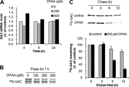
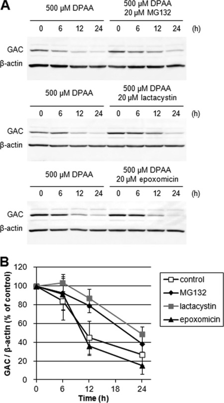
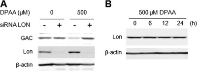
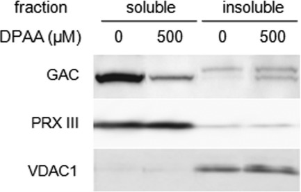
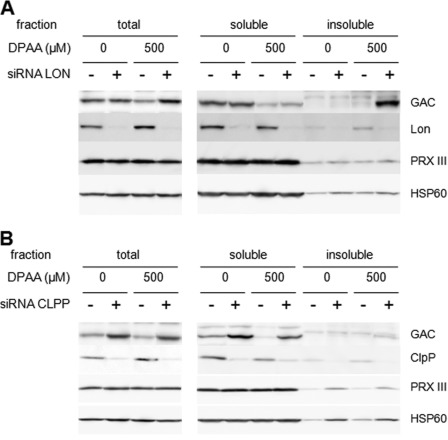
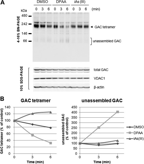
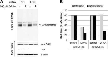
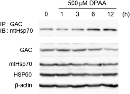
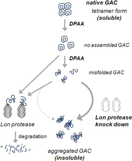
Glutaminase C (GAC), a splicing variant of the kidney-type glutaminase (KGA) gene, is a vital mitochondrial enzyme protein that catalyzes glutamine to glutamate. Earlier studies have shown that GAC proteins in the human hepatocarcinoma cell line, HepG2, were down-regulated by diphenylarsinic acid (DPAA), but the mechanism by which DPAA induced GAC protein down-regulation remained poorly understood. Here, we showed that DPAA promoted GAC protein degradation without affecting GAC transcription and translation. Moreover, DPAA-induced GAC proteolysis was mediated by mitochondrial Lon protease. DPAA insolubilized 0.5% Triton X-100-soluble GAC protein and promoted the accumulation of insoluble GAC in Lon protease knockdown cells. DPAA destroyed the native tetrameric GAC conformation and promoted an increase in the unassembled form of GAC when DPAA was incubated with cell extracts. Decreases in the tetrameric form of GAC were observed in cells exposed to DPAA, and decreases occurred prior to a decrease in total GAC protein levels. In addition, decreases in the tetrameric form of GAC were observed independently with Lon protease. Mitochondrial heat shock protein 70 is known to be an indispensable protein that can bind to misfolded proteins, thereby supporting degradation of proteins sensitive to Lon protease. When cells were incubated with DPAA, GAC proteins that can bind with mtHsp70 increased. Interestingly, the association of mtHsp70 with GAC protein increased when the tetrameric form of GAC was reduced. These results suggest that degradation of native tetrameric GAC by DPAA may be a trigger in GAC protein degradation by Lon protease.


Figures

FIGURE 1.
FIGURE 1.
Effects of DPAA on GAC mRNA expression, translation, and GAC proteolysis. A, GAC mRNA expression levels after treatment with DPAA. GAC mRNA levels were analyzed by quantitative real time PCR in HepG2 cells that were incubated with 0, 250, and 500 μm DPAA for 4, 8, and 24 h. The reproducibility of results was confirmed by separate experiments. B, newly translated GAC protein levels after treatment with DPAA. HepG2 cells were cultured for 24 h in 0–500 μm DPAA-containing culture medium, which was then replaced with [35S]methionine-containing medium and pulsed for 1 h, followed by immunoprecipitation of GAC, PAGE, and autoradiography. The reproducibility of results was confirmed by separate experiments. C, proteolysis of GAC protein by DPAA. Upper panel, HepG2 cells were pulsed for 2 h with [35S]methionine and chased at the indicated times with or without 500 μm DPAA-containing chase medium, followed by immunoprecipitation of GAC, PAGE, and autoradiography. Lower panel, a quantitative phosphoimager analysis of results. Data are presented as the percentage of labeling at time 0 of the chase (mean ± S.D., n = 3).
FIGURE 2.
FIGURE 2.
Effects of 26 S proteasome inhibitors on DPAA-induced GAC degradation. A, immunoblot pattern of GAC expression levels in HepG2 cells after treatment by DPAA with or without proteasome inhibitor. HepG2 cells were preincubated for 2 h in each proteasome inhibitor (lactacystin, MG132, and epoxomicin)-containing medium, after which DPAA was added and the culture continued for the indicated time. A cell extract was prepared, and immunoblot analysis was performed as described under “Experimental Procedures.” B, quantitative analysis of GAC protein levels. GAC protein levels are presented as the percentage of levels at 0 h (mean ± S.D., n = 3).
FIGURE 3.
FIGURE 3.
Effect of Lon protease on DPAA-induced GAC degradation. A, GAC protein levels in Lon protease knockdown cells after treatment with or without DPAA. Double-stranded oligonucleotides for Lon protease (siRNA LON +) or negative control oligonucleotides (siRNA LON −) were transiently transfected into HepG2 cells for 48 h, and then the medium was replaced with fresh medium. After 40 h of culture, 500 μm DPAA was added, and incubation was continued for 12 h. Cellular GAC and Lon protease protein levels were determined by immunoblot analysis as described under “Experimental Procedures.” The reproducibility of results was confirmed by separate experiments. B, changes in Lon protease protein levels after treatment with DPAA. HepG2 cells were cultured in medium containing 500 μm DPAA for up to 24 h, and Lon protease levels were examined by immunoblot analysis. β-Actin was indicated as the loading control. The reproducibility of results was confirmed by separate experiments.
FIGURE 4.
FIGURE 4.
Changes in the subcellular distribution of GAC proteins after treatment with DPAA. After treatment with 500 μm DPAA for 12 h, soluble and insoluble cellular proteins in HepG2 cells were prepared as described under “Experimental Procedures.” Contamination of the soluble or insoluble membrane fractions was checked by immunoblots using antibodies against PRX III, which is a mitochondrial soluble matrix protein, and voltage-dependent anion channel-1 (VDAC1), which is a component of porin that is embedded in the mitochondrial outer membrane. The reproducibility of results was confirmed by separate experiments.
FIGURE 5.
FIGURE 5.
Subcellular distribution of GAC protein in DPAA-treated Lon protease or ClpP protease knockdown cells. A, subcellular distribution of GAC protein in Lon protease knockdown cells after treatment with DPAA. Double-stranded oligonucleotides for Lon protease (siRNA LON +) or negative control oligonucleotides (siRNA LON −) were transiently transfected into HepG2 cells for 48 h and then the medium was replaced with fresh medium. After a 40-h incubation, 500 μm DPAA was added and incubated further for 12 h. Total, soluble, and insoluble cellular extracts of HepG2 cells were prepared as described under “Experimental Procedures.” GAC and other mitochondrial protein (PRX III and HSP60) levels in each fraction were determined by immunoblot analysis as described under “Experimental Procedures.” The reproducibility of results was confirmed by separate experiments. B, subcellular distribution of GAC protein in ClpP protease knockdown cells after treatment with DPAA. Double-stranded oligonucleotides for ClpP protease (siRNA CLPP +) or negative control oligonucleotides (siRNA CLPP −) were transiently transfected into HepG2 cells for 48 h and then the medium was replaced with fresh medium. After a 40-h incubation, 500 μm DPAA was added and incubated further for 12 h. Total, soluble, and insoluble cellular extracts of HepG2 cells were prepared as described under “Experimental Procedures.” GAC and other mitochondrial protein (PRX III and HSP60) levels in each fraction were determined by immunoblot analysis as described under “Experimental Procedures.” The reproducibility of results was confirmed by separate experiments.
FIGURE 6.
FIGURE 6.
DPAA directly destroys the native tetrameric GAC and forms unassembled GAC. A, BN-PAGE analysis of native GAC protein after incubation with DPAA. HepG2 cell extracts were prepared and incubated with 14.8 mm DPAA or NaAsO2 for the indicated time points at 25 °C. Native GAC proteins were determined with 4–16% BN-PAGE and immunoblot analysis as described under “Experimental Procedures.” Total GAC, VDAC1, and β-actin levels in cell extracts were determined by SDS-PAGE and immunoblot analysis. B, changes in tetrameric and unassembled forms of GAC after incubation with DPAA. Each form of GAC protein prior to incubation at 25 °C was set to 100%.
FIGURE 7.
FIGURE 7.
Native tetrameric GAC in HepG2 cells treated with DPAA rapidly disappears before total cellular GAC degradation. A, BN-PAGE analysis of native GAC protein in cells treated with DPAA. HepG2 cells were incubated with 500 μm DPAA or 25 μm NaAsO2 for the indicated time points, and native tetrameric GAC levels were determined with 4–16% BN-PAGE and immunoblot analysis as described under “Experimental Procedures.” Total GAC, VDAC1, and β-actin levels in cell extracts were determined by SDS-PAGE and immunoblot analysis. B, quantitative analysis of total and tetrameric GAC protein levels in HepG2 cells treated with DPAA. GAC levels in cells cultured without DPAA medium were set to 100%.
FIGURE 8.
FIGURE 8.
DPAA promotes native tetrameric GAC destruction in both control and Lon protease knockdown cells. A, tetrameric GAC levels in Lon protease knockdown cells before and after treatment with DPAA. Double-stranded oligonucleotides for Lon protease (siRNA LON) or negative control oligonucleotides (siRNA NC) were transiently transfected into HepG2 cells for 48 h, and then the medium was replaced with fresh medium. After a 40-h incubation, 500 μm DPAA was added and incubated for 12 h. Native tetrameric GAC was determined by BN-PAGE and immunoblot analysis. Total GAC, Lon, and β-actin levels were determined by SDS-PAGE and immunoblot analysis as described under “Experimental Procedures.” B, quantity of total and tetrameric GAC protein levels in Lon protease knockdown HepG2 cells treated with DPAA. GAC levels in cells cultured without DPAA medium were set to 100%.
FIGURE 9.
FIGURE 9.
PDAA promotes the interaction of GAC with mtHsp70. HepG2 cells were incubated with DPAA for the indicated time points, and immunoprecipitation (IP) was performed as described under “Experimental Procedures.” Each protein level in cell extracts and immunoprecipitants was determined by SDS-PAGE and immunoblot (IB) analysis.
FIGURE 10.
FIGURE 10.
Hypothetical illustration of DPAA-induced GAC protein degradation. In the normal state, GAC proteins assembled each other and formed the soluble tetrameric complex. In the presence of DPAA, tetrameric GAC was destroyed, and unassembled GAC was formed. It is obscure whether DPAA promoted unassembled GAC modifications; however, unassembled GAC itself or further modified ones may be degraded by mitochondrial Lon protease. If functional Lon protease was lost, GAC proteins may accumulate and form an insoluble aggregate.

Similar articles

Cited by

References

    1. Ishii K., Tamaoka A., Otsuka F., Iwasaki N., Shin K., Matsui A., Endo G., Kumagai Y., Ishii T., Shoji S., Ogata T., Ishizaki M., Doi M., Shimojo N. (2004) Diphenylarsinic acid poisoning from chemical weapons in Kamisu, Japan. Ann. Neurol. 56, 741–745 - PubMed
    1. Kita K., Suzuki T., Ochi T. (2007) Down-regulation of glutaminase C in human hepatocarcinoma cell by diphenylarsinic acid, a degradation product of chemical warfare agents. Toxicol. Appl. Pharmacol. 220, 262–270 - PubMed
    1. Curthoys N. P., Watford M. (1995) Regulation of glutaminase activity and glutamine metabolism. Annu. Rev. Nutr. 15, 133–159 - PubMed
    1. Elgadi K. M., Meguid R. A., Qian M., Souba W. W., Abcouwer S. F. (1999) Cloning and analysis of unique human glutaminase isoforms generated by tissue-specific alternative splicing. Physiol. Genomics 1, 51–62 - PubMed
    1. Holcomb T., Taylor L., Trohkimoinen J., Curthoys N. P. (2000) Isolation, characterization, and expression of a human brain mitochondrial glutaminase cDNA. Mol. Brain Res. 76, 56–63 - PubMed

Publication types

LinkOut - more resources