. 2012 May 25;287(22):18535-43.
doi: 10.1074/jbc.M112.357327. Epub 2012 Apr 5.

Functional asymmetries of proteasome translocase pore

Affiliations

Functional asymmetries of proteasome translocase pore

Jenny Erales et al. J Biol Chem. .

Abstract

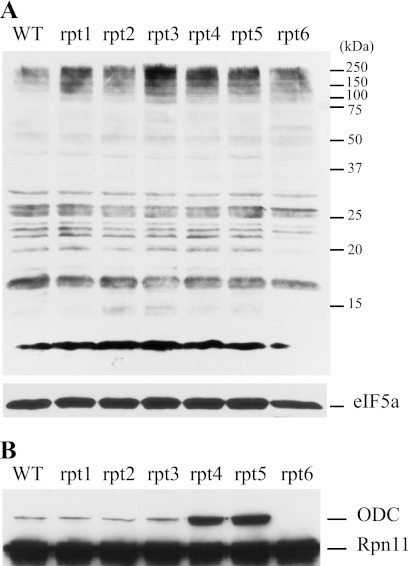
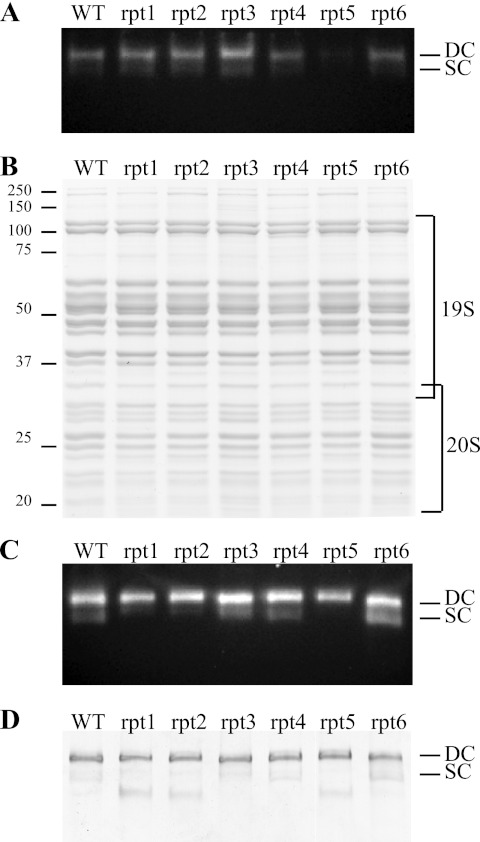
Degradation by proteasomes involves coupled translocation and unfolding of its protein substrates. Six distinct but paralogous proteasome ATPase proteins, Rpt1 to -6, form a heterohexameric ring that acts on substrates. An axially positioned loop (Ar-Φ loop) moves in concert with ATP hydrolysis, engages substrate, and propels it into a proteolytic chamber. The aromatic (Ar) residue of the Ar-Φ loop in all six Rpts of S. cerevisiae is tyrosine; this amino acid is thought to have important functional contacts with substrate. Six yeast strains were constructed and characterized in which Tyr was individually mutated to Ala. The mutant cells were viable and had distinct phenotypes. rpt3, rpt4, and rpt5 Tyr/Ala mutants, which cluster on one side of the ATPase hexamer, were substantially impaired in their capacity to degrade substrates. In contrast, rpt1, rpt2, and rpt6 mutants equaled or exceeded wild type in degradation activity. However, rpt1 and rpt6 mutants had defects that limited cell growth or viability under conditions that stressed the ubiquitin proteasome system. In contrast, the rpt3 mutant grew faster than wild type and to a smaller size, a defect that has previously been associated with misregulation of G1 cyclins. This rpt3 phenotype probably results from altered degradation of cell cycle regulatory proteins. Finally, mutation of five of the Rpt subunits increased proteasome ATPase activity, implying bidirectional coupling between the Ar-Φ loop and the ATP hydrolysis site. The present observations assign specific functions to individual Rpt proteins and provide insights into the diverse roles of the axial loops of individual proteasome ATPases.


Figures

FIGURE 1.
FIGURE 1.
Yeast growth. A, serial 10-fold dilutions of liquid cultures were spotted onto plates with various media and incubated as indicated. Can and CHX, canavanine and cycloheximide (0.5 μg/ml), respectively. B, growth of wild type or mutant rpt strains at 30 °C in YPD was followed by measuring culture turbidity at 600 nm. C, size distribution of wild type and rpt3 strains. Samples were removed from the cultures shown in B at 8 or 30 h, and the distribution of cell diameters was determined by a Coulter Counter.
FIGURE 2.
FIGURE 2.
Impairment of degradation in vivo. Shown is accumulation of polyubiquitinated protein (A) and FLAG-ODC (B). Lysates were prepared from the indicated strains, separated by SDS-PAGE on a 12% gel, and analyzed by immunoblotting with an anti-ubiquitin antibody (or with antibody to eIF5a as loading control) (A) or anti-FLAG (B). Anti-FLAG recognize both FLAG-ODC and Rpn11-FLAG proteins.
FIGURE 3.
FIGURE 3.
Proteasome integrity, composition, and function. A, proteasome integrity after subcellular fractionation. Lysates were prepared from the indicated strains, and 50 μg were loaded on a 4% native gel. Proteasomes were visualized by incubating the gel with N-succinyl-LLVY-7-amino-4-methylcoumarin peptide, a fluorogenic proteasome substrate. B, 12% SDS-PAGE and Coomassie staining of 6 μg of purified wild type and mutated proteasomes. C and D, 4% native gel analysis of 2 μg of purified wild type and mutated proteasomes, revealed by peptidase activity (C) or by Coomassie staining (D). DC and SC, doubly capped and singly capped proteasomes, respectively.
FIGURE 4.
FIGURE 4.
ATPase activity of the proteasome. ATPase activity was measured in the presence of 50 nm proteasome and in excess of ATP (5 mm). The catalytic constant (kcat) is the mean of three or four experiments. Error bars, S.D.
FIGURE 5.
FIGURE 5.
In vitro degradation of substrates. Initial rates were determined for 9 min in the presence of 50 nm proteasome and were initiated with 1 μm substrate, either R-I27-ext or R-I27V13P-ext. The activity of the mutant proteasomes is represented as a percentage compared with the wild type proteasomes for each substrate. For wild type proteasomes, the value of the catalytic constant (min−1) was 0.036 ± 0.001 and 0.159 ± 0.008 for R-I27-ext and R-I27V13P-ext, respectively. Error bars, S.D. of four independent experiments.

Similar articles

Cited by

References

    1. Glickman M. H., Rubin D. M., Fried V. A., Finley D. (1998) The regulatory particle of the Saccharomyces cerevisiae proteasome. Mol. Cell Biol. 18, 3149–3162 - PMC - PubMed
    1. Groll M., Ditzel L., Löwe J., Stock D., Bochtler M., Bartunik H. D., Huber R. (1997) Structure of 20S proteasome from yeast at 2.4 Å resolution. Nature 386, 463–471 - PubMed
    1. Glickman M. H., Rubin D. M., Coux O., Wefes I., Pfeifer G., Cjeka Z., Baumeister W., Fried V. A., Finley D. (1998) A subcomplex of the proteasome regulatory particle required for ubiquitin-conjugate degradation and related to the COP9-signalosome and eIF3. Cell 94, 615–623 - PubMed
    1. Tomko R. J., Jr., Funakoshi M., Schneider K., Wang J., Hochstrasser M. (2010) Heterohexameric ring arrangement of the eukaryotic proteasomal ATPases. Implications for proteasome structure and assembly. Mol. Cell 38, 393–403 - PMC - PubMed
    1. Lasker K., Förster F., Bohn S., Walzthoeni T., Villa E., Unverdorben P., Beck F., Aebersold R., Sali A., Baumeister W. (2012) Molecular architecture of the 26S proteasome holocomplex determined by an integrative approach. Proc. Natl. Acad. Sci. U.S.A. 109, 1380–1387 - PMC - PubMed

Publication types

MeSH terms

Substances