. 2012 May 25;287(22):17975-84.
doi: 10.1074/jbc.M111.310458. Epub 2012 Apr 6.

α6β4 integrin, a master regulator of expression of integrins in human keratinocytes

Affiliations

α6β4 integrin, a master regulator of expression of integrins in human keratinocytes

Kristina R Kligys et al. J Biol Chem. .

Abstract

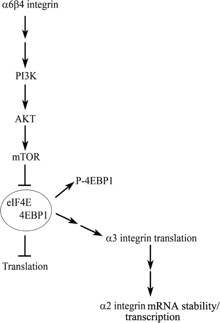
Three major laminin and collagen-binding integrins in skin (α6β4, α3β1, and α2β1) are involved in keratinocyte adhesion to the dermis and dissemination of skin cells during wound healing and/or tumorigenesis. Knockdown of α6 integrin in keratinocytes not only results in motility defects but also leads to decreased surface expression of the α2, α3, and β4 integrin subunits. Whereas α2 integrin mRNA levels are decreased in α6 integrin knockdown cells, α3 and β4 integrin mRNAs levels are unaffected. Expression of either α6 or α3 integrin in α6 integrin knockdown cells restores α2 integrin mRNA levels. Moreover, re-expression of α6 integrin increases β4 integrin protein at the cell surface, which results in an increase in α3 integrin expression via activation of initiation factor 4E-binding protein 1. Our data indicate that the α6β4 integrin is a master regulator of transcription and translation of other integrin subunits and underscore its pivotal role in wound healing and cancer.


Figures

FIGURE 1.
FIGURE 1.
Stable knockdown of α6 integrin expression in human keratinocytes. Human keratinocytes were infected with lentivirus encoding α6 integrin shRNA and stably selected for the loss of α6 integrin expression. α6 and β4 integrin expression was measured in three knockdown clones (1–3, as indicated) by quantitative RT-PCR (A). The bars of the graph represent the relative expression (±S.D.) of α6 and β4 integrin mRNA in the shRNA clones normalized to GAPDH and compared with HEK. Samples were measured in triplicate for two individual experiments. B, α6 integrin, β4 integrin, actin, BP180, mTOR, and lamin were evaluated in the same clones by immunoblot. β-Actin, mTOR, and lamin were used as loading controls. C, α6 and β4 integrin localization was determined by confocal immunofluorescence (α6 shRNA clone 1 only; bar, 10 μm). D, surface expression of α6 and β4 integrins in the three knockdown clones was evaluated by FACS (top two panels). The black curves represent secondary antibody alone. In the lower two panels of D and E, wild-type cells, α6 shRNA clone 1 cells, and α6 shRNA clone 1 cells infected with adenovirus encoding a refractory α6 integrin construct to re-express α6 integrin (+refα6) were analyzed by FACS and immunoblot. In E, the graph represents the relative level of β4 integrin protein expression in cells compared with parental HEK, quantified from immunoblots as shown to the left. The individual bars represent the means ± S.E. (n = 3). The p values were generated by Student's t test. **, p ≤ 0.001. The results using control (wild-type) keratinocytes (HEK) are presented in each assay.
FIGURE 2.
FIGURE 2.
Loss of α6β4 integrin leads to decreased α2 and α3 integrin expression. α2 and α3 integrin expression was measured in control cells (HEK), α6 shRNA clones 1–3, and α6 shRNA clone 1 cells induced to re-express GFP-tagged α6 integrin (+refα6) by FACS (A, B, F, and G) and quantitative RT-PCR (C–E). The individual bars on the graphs represent the relative levels of α2 or α3 integrin mRNA normalized to GAPDH and compared with HEK (±S.D.). The samples were measured in triplicate in two experiments. In E, α2 integrin mRNA expression was also measured in α6 shRNA clone 1 keratinocytes infected with adenovirus expressing α3 integrin (+α3). In H, α3 integrin surface expression was measured by FACS analysis in β4 integrin-deficient keratinocytes (JEB) and JEB cells re-expressing β4 integrin. The black curves on the FACS analyses represent secondary antibody alone.
FIGURE 3.
FIGURE 3.
Human keratinocytes lacking α6β4 integrin expression display adhesion and migration defects. In A, control keratinocytes (HEK) and α6 shRNA clone 1 cells were assayed for adhesion to laminin-332- or collagen I-coated dishes at 1 h after plating. The number of adherent cells was measured by reading the absorbance at 570 nm. The average absorbance for HEK was taken as 100%. The individual bars on the graph represent the means ± S.E. (n = 3) with samples measured in triplicate for each experiment. p values were derived by comparing samples to HEK (Student's t test). *, p ≤ 0.05. B, vector diagrams depict the individual migration patterns of wild-type keratinocytes (HEK) (n = 11), α6 shRNA clone 1 cells (n = 12), or α6 shRNA clone 1 cells induced to re-express GFP-tagged α6 integrin (+refα6) (n = 12). The cells were plated on glass and tracked over a 2-h period. C, Rac1 activity in control HEK and α6 shRNA clone 1 cells was measured by the G-LISA Rac1 activation assay. The individual bars of the graph represent the means ± S.E. (n = 3) (in each experiment, duplicate samples were assayed). p value was derived by comparing to HEK (Student's t test). *, p ≤ 0.05. D, graph representing the average velocity of cells plated on glass over a 2-h period. The individual bars of the graph represent the means ± S.E. (n ≥ 50 cells). **, p ≤ 0.001. E, vector diagrams depicting the migration pattern of HEK (n = 28), α6 shRNA clone 1 cells (n = 37), or α6 shRNA clone 1 cells infected with retrovirus encoding GFP-tagged α3 integrin (+α3) plated on matrix deposited by HEK (n = 19). The cells were allowed to adhere to the matrix for 2 h, and the migration of cells was subsequently tracked for 2 h. F, graph represents the average migration index (net displacement/total distance) of cells over a 2-h period. The individual bars of the graph represent the means ± S.E. (n ≥ 50 cells). G, graph represents the average velocity of cells plated on pre-formed matrix over a 2-h period. The individual bars of the graph represent the means ± S.E. (n ≥ 50 cells). p values were derived by Student's t test. *, p ≤ 0.05.
FIGURE 4.
FIGURE 4.
α6β4 integrin regulates the translation of α3 integrin through 4EBP1. A, polysome fractions from control keratinocytes or α6 shRNA clone 1 keratinocytes were isolated, and the levels of α3 integrin (left), β4 integrin (middle), and GAPDH (right) mRNA were determined by quantitative RT-PCR, as indicated. The graphs represent the relative levels of α3, β4, or GAPDH mRNA normalized to the ribosomal protein S26 and compared with HEK. The samples were measured in triplicate, and the graphs represent the means ± S.E. of three independent experiments. B, whole cell extracts from wild-type (HEK), α6 shRNA clone 1 cells, or α6 shRNA cone 1 cells induced to re-express α6 integrin (+refα6) were probed for levels of phosphorylated 4EBP1 and total 4EBP1. C and D, whole cell extracts from wild-type (HEK), α6 shRNA clone 1 cells, or α6 shRNA clone 1 cells infected with adenovirus encoding myristoylated AKT were probed for levels of phosphorylated 4EBP1 and total 4EBP1 (C) or β4 integrin and α3 integrin (D). Actin and lamin A/C were used as loading controls in B–D. Graphs in B–D represent the means expression (normalized to lamin A/C) + S.E. of three independent experiments, quantified from immunoblots.
FIGURE 5.
FIGURE 5.
Schematic of α6β4 integrin regulation of α3 and α2 integrin expression. α6β4 integrin signaling to PI3K leads to activation of AKT and its downstream effectors. Activation of this pathway leads to phosphorylation/inactivation of 4EBP1, dissociation of the complex containing initiation factor 4E (eIF4E) and 4EBP1, and ultimately, the translation of α3 integrin. Expression of α3 integrin in turn mediates the expression of α2 integrin by regulating the transcription of the α2 integrin gene and/or α2 integrin mRNA stability.

References

    1. DiPersio C. M., van der Neut R., Georges-Labouesse E., Kreidberg J. A., Sonnenberg A., Hynes R. O. (2000) α3β1 and α6β4 integrin receptors for laminin-5 are not essential for epidermal morphogenesis and homeostasis during skin development. J. Cell Sci. 113, 3051–3062 - PubMed
    1. Dowling J., Yu Q. C., Fuchs E. (1996) β4 integrin is required for hemidesmosome formation, cell adhesion and cell survival. J. Cell Biol. 134, 559–572 - PMC - PubMed
    1. Georges-Labouesse E., Messaddeq N., Yehia G., Cadalbert L., Dierich A., Le Meur M. (1996) Absence of integrin α6 leads to epidermolysis bullosa and neonatal death in mice. Nat. Genet. 13, 370–373 - PubMed
    1. Brakebusch C., Grose R., Quondamatteo F., Ramirez A., Jorcano J. L., Pirro A., Svensson M., Herken R., Sasaki T., Timpl R., Werner S., Fässler R. (2000) Skin and hair follicle integrity is crucially dependent on β1 integrin expression on keratinocytes. EMBO J. 19, 3990–4003 - PMC - PubMed
    1. van der Neut R., Krimpenfort P., Calafat J., Niessen C. M., Sonnenberg A. (1996) Epithelial detachment due to absence of hemidesmosomes in integrin β4 null mice. Nat. Genet. 13, 366–369 - PubMed

Publication types