. 2012 May 25;287(22):18492-9.
doi: 10.1074/jbc.M112.351023. Epub 2012 Apr 6.

The cytoskeletal protein α-catenin unfurls upon binding to vinculin

Affiliations

The cytoskeletal protein α-catenin unfurls upon binding to vinculin

Erumbi S Rangarajan et al. J Biol Chem. .

Abstract

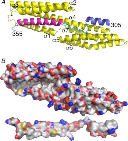
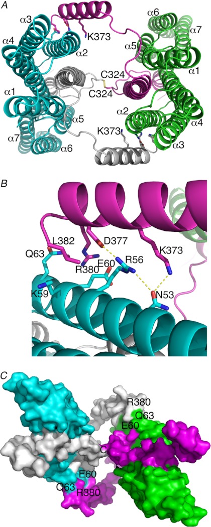
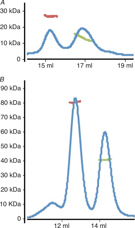
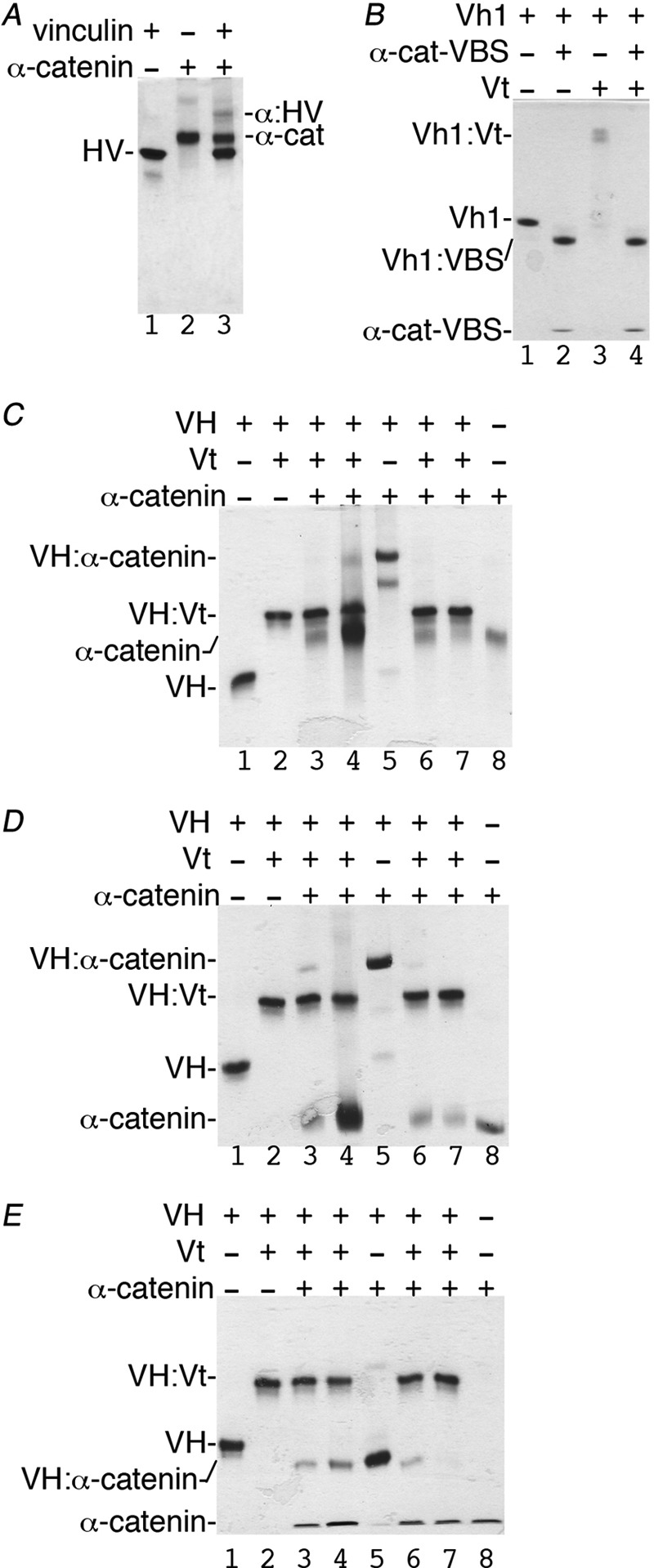
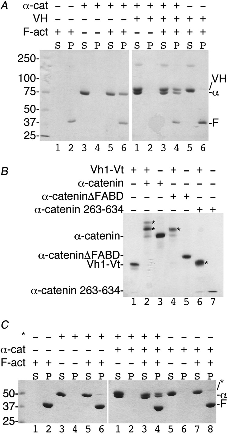
Adherens junctions (AJs) are essential for cell-cell contacts, morphogenesis, and the development of all higher eukaryotes. AJs are formed by calcium-dependent homotypic interactions of the ectodomains of single membrane-pass cadherin family receptors. These homotypic interactions in turn promote binding of the intracellular cytoplasmic tail domains of cadherin receptors with β-catenin, a multifunctional protein that plays roles in both transcription and AJs. The cadherin receptor-β-catenin complex binds to the cytoskeletal protein α-catenin, which is essential for both the formation and the stabilization of these junctions. Precisely how α-catenin contributes to the formation and stabilization of AJs is hotly debated, although the latter is thought to involve its interactions with the cytoskeletal protein vinculin. Here we report the crystal structure of the vinculin binding domain (VBD) of α-catenin in complex with the vinculin head domain (Vh1). This structure reveals that α-catenin is in a unique unfurled mode allowing dimer formation when bound to vinculin. Finally, binding studies suggest that vinculin must be in an activated state to bind to α-catenin and that this interaction is stabilized by the formation of a ternary α-catenin-vinculin-F-actin complex, which can be formed via the F-actin binding domain of either protein. We propose a feed-forward model whereby α-catenin-vinculin interactions promote their binding to the actin cytoskeleton to stabilize AJs.


Figures

FIGURE 1.
FIGURE 1.
α-Catenin is unfurled when bound to vinculin. A, the crystal structure of Vh1 (residues 1–258; yellow) in complex with α-catenin VBD shows an extended conformation of α-catenin with extensive interactions with both subdomains of Vh1. Vh1 α-helices and α-catenin termini are labeled. Residues 305–316 are shown in blue, 328–351 are in magenta, and the remainder of the α-catenin residues are in cyan. For clarity, residues 290–304 and 362–382 are not shown. B, surface representation showing the respective interfaces (white, carbon; red, oxygen; blue, nitrogen; yellow, sulfur). Vh1 is in the same orientation as shown in A, and α-catenin is rotated 180° down with respect to its orientation shown in A.
FIGURE 2.
FIGURE 2.
α-Catenin-vinculin complex crystal structure. Vh1 is shown in cyan or green, and its respective bound α-catenin is shown in white or magenta. A, α-catenin Cys-324 forms a disulfide bond with a two-fold related molecule. Likewise, the C-terminal α-catenin α-helix binds to the second Vh1 α-helix of a two-fold related heterodimer burying over 500 Å2 of solvent-accessible surface area with a shape correlation statistic derived using the CCP4 program SC (55) of over 0.8 for this interface, a significant value where a value of 1 indicates a perfect fit versus 0.35, which indicates the mismatch of an artificial association. Vh1 α-helices are labeled, as well as some α-catenin residues 324 and 373. B, close-up view of the dimer interface. Vinculin residues are shown in cyan, and α-catenin are in magenta as in A and in a slightly rotated view from A. C, space-filling representation of the dimer of heterodimers (same color coding as shown in A) and rotated about 180° about the horizontal axis of the A image. Some contact residues are indicated.
FIGURE 3.
FIGURE 3.
VBD alone or in complex with Vh1 is a monomer and dimer in solution. A and B, the oligomeric states of the α-catenin VBD either alone (A) or in complex with vinculin (B) are estimated by multiangle light scattering combined with size exclusion chromatography, and the respective profiles are provided. The blue line represents the UV absorbance (without units), whereas the green and red lines denote the molecular masses (shown in kDa on the ordinate) as calculated by ASTRA (software provided by Wyatt Technology) as follows: A, 13.6 and 27.2 kDa where the molecular mass of VBD is 12.3 kDa; B, 40.9 and 80.5 kDa where the molecular mass of the 1:1 Vh1-VBD complex is 41.1 kDa.
FIGURE 4.
FIGURE 4.
Vt has higher affinity for VH when compared with α-catenin. A, native gel shift assay shows that some full-length human vinculin (HV, residues 1–1066, lane 1) binds to full-length α-catenin (α-cat, residues 82–906, lane 2; complex, α-HV, lane 3). B, the minimal α-catenin VBS (α-cat:VBS; residues 332–353) displaces Vt from the Vh1-Vt complex (lane 3) to form a new Vh1-α-catenin-VBS complex (Vh1:VBS, lanes 2 and 4) that migrates farther than Vh1 alone (lane 1). C–E, reciprocal competition assays by titrating equimolar concentrations (15 μm) or 5-fold excess concentrations (75 μm) of α-catenin (lanes 3 and 4) residues 82–906 (C), residues 82–634 (D), or 263–634 (E) to VH-Vt versus titration of increasing amounts of Vt to VH in complex with α-catenin (lanes 6 and 7) residues 82–906 (C), 82–634 (D), or 263–634 (E) as analyzed by native gel shift mobility assays. VH alone (lane 1), VH-Vt (lane 2), VH-α-catenin (lane 5), and α-catenin (lane 8) are also shown. As is evident, all three α-catenin constructs fail to displace Vt from preformed VH-Vt complexes (lanes 3 and 4). In contrast, Vt readily displaces all three α-catenin constructs from preformed VH-α-catenin complexes (lanes 6 and 7). VH, vinculin head domain (residues 1–843); Vt, vinculin tail domain (residues 879–1066).
FIGURE 5.
FIGURE 5.
Vinculin-α-catenin complex co-sediments with F-actin via F-actin binding domain (FABD) of either α-catenin or vinculin. A, the α-catenin-VH complex binds F-actin via the α-catenin FABD. About 30 μm of either vinculin or α-catenin was used in the corresponding reaction mixtures. Left gel, lanes 1 and 2, F-actin is found in the pellet (P). Lanes 3 and 4, α-catenin does not aggregate and remains in the soluble fraction (S). Lanes 5 and 6, α-catenin binds to F-actin. Right gel, lanes 1 and 2, the VH-α-catenin complex does not aggregate. Lanes 3 and 4, the VH-α-catenin complex binds to F-actin via the α-catenin FABD. Lanes 5 and 6, VH does not bind to F-actin. Molecular weight markers are shown for the left gel. α-cat or α, α-catenin (residues 144–906); VH, vinculin head domain (residues 1–843); F-act or F, F-actin. B, the Vt2 domain seems to hinder closed, inactive vinculin to bind to α-catenin. The addition of α-catenin (residues 82–906, 20 μm, lane 2), α-cateninΔFABD (residues 82–634, 20 μm, lane 4), or α-catenin 263–634 (20 μm, lane 6) to the Vh1-Vt chimera (residues 1–258 and 848–1066, 15 μm) lacking the Vt2 domain results in α-catenin-vinculin complex formation (indicated by asterisks). Binding in each case was saturable with no unbound α-catenin remaining. C, the α-cateninΔFABD-vinculin complex binds F-actin via Vt. Left gel, lanes 1 and 2, F-actin is found in the pellet (P). Lanes 3 and 4, the Vh1-Vt chimera (asterisks) does not aggregate and remains in the soluble fraction (S). Lanes 5 and 6, the Vh1-Vt chimera does not bind to F-actin. Right gel, lanes 1 and 2, the Vh1-Vt chimera in complex with α-cateninΔFABD does not aggregate. Lanes 3 and 4, the Vh1-Vt chimera in complex with α-cateninΔFABD binds F-actin via Vt. Lanes 5 and 6), α-cateninΔFABD does not aggregate. Lanes 7 and 8, α-cateninΔFABD does not bind to F-actin. α-cat or α, α-catenin (residues 144–906); F-act or F, F-actin. Molecular weight markers are shown for the left gel.

References

    1. Braga V. M., Balda M. S. (2004) Regulation of cell-cell adhesion. Semin. Cell Dev. Biol. 15, 631–632 - PubMed
    1. Pokutta S., Weis W. I. (2002) The cytoplasmic face of cell contact sites. Curr. Opin. Struct. Biol. 12, 255–262 - PubMed
    1. Clevers H. (2006) Wnt/β-Catenin signaling in development and disease. Cell 127, 469–480 - PubMed
    1. Wahl J. K., 3rd, Kim Y. J., Cullen J. M., Johnson K. R., Wheelock M. J. (2003) N-cadherin-catenin complexes form prior to cleavage of the proregion and transport to the plasma membrane. J. Biol. Chem. 278, 17269–17276 - PubMed
    1. Haegel H., Larue L., Ohsugi M., Fedorov L., Herrenknecht K., Kemler R. (1995) Lack of β-catenin affects mouse development at gastrulation. Development 121, 3529–3537 - PubMed

Publication types

Associated data