. 2012 May 25;287(22):18351-8.
doi: 10.1074/jbc.M112.350918. Epub 2012 Apr 9.

Roles for peroxisome proliferator-activated receptor γ (PPARγ) and PPARγ coactivators 1α and 1β in regulating response of white and brown adipocytes to hypoxia

Affiliations

Roles for peroxisome proliferator-activated receptor γ (PPARγ) and PPARγ coactivators 1α and 1β in regulating response of white and brown adipocytes to hypoxia

Elizabeth Pino et al. J Biol Chem. .

Abstract

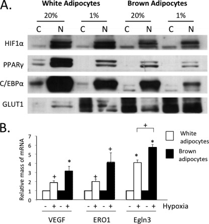
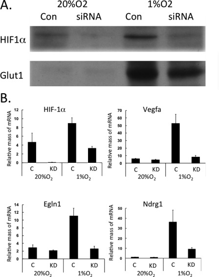
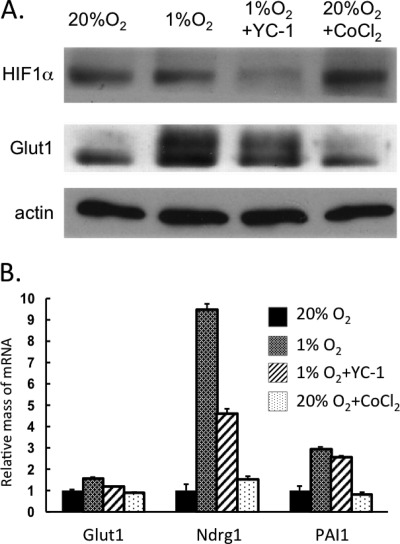
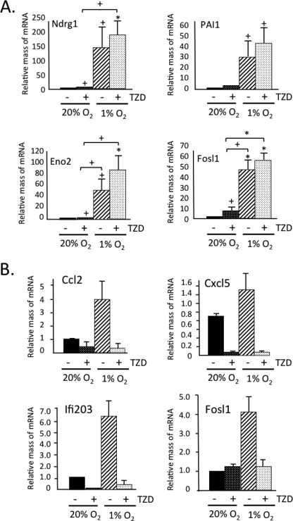
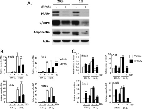
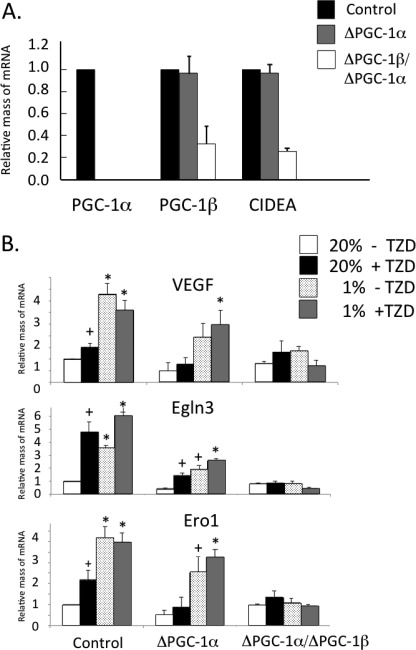
Obese white adipose tissue is hypoxic but is incapable of inducing compensatory angiogenesis. Brown adipose tissue is highly vascularized, facilitating delivery of nutrients to brown adipocytes for heat production. In this study, we investigated the mechanisms by which white and brown adipocytes respond to hypoxia. Brown adipocytes produced lower amounts of hypoxia-inducible factor 1α (HIF-1α) than white adipocytes in response to low O(2) but induced higher levels of hypoxia-associated genes. The response of white adipocytes to hypoxia required HIF-1α, but its presence alone was incapable of inducing target gene expression under normoxic conditions. In addition to the HIF-1α targets, hypoxia also induced many inflammatory genes. Exposure of white adipocytes to a peroxisome proliferator-activated receptor γ (PPARγ) ligand (troglitazone) attenuated induction of these genes but enhanced expression of the HIF-1α targets. Knockdown of PPARγ in mature white adipocytes prevented the usual robust induction of HIF-1α targets in response to hypoxia. Similarly, knockdown of PPARγ coactivator (PGC) 1β in PGC-1α-deficient brown adipocytes eliminated their response to hypoxia. These data demonstrate that the response of white adipocytes requires HIF-1α but also depends on PPARγ in white cells and the PPARγ cofactors PGC-1α and PGC-1β in brown cells.


Figures

FIGURE 1.
FIGURE 1.
In response to hypoxia, brown adipocytes produce lower amounts of HIF-1α than white adipocytes but induce higher levels of hypoxia-associated genes. 3T3-L1 and brown preadipocytes were differentiated until day 7, at which stage cells were exposed to 20% or 1% O2 for 18 h. A, nuclear (N) and cytoplasmic (C) cell fractions were prepared, and extracts were subjected to Western blot analysis. B, cells were also harvested for RNA isolation and quantitative real-time PCR analysis. Results are means ± S.E. (n = 3). *, p < 0.01; +, p < 0.05. C/EBPα, CCAAT/enhancer-binding protein α.
FIGURE 2.
FIGURE 2.
Response of white adipocytes to hypoxia requires HIF-1α. 3T3-L1 preadipocytes were differentiated until day 8, at which time cells were exposed to DeliverX siRNA reagent alone (C, Con) or with oligonucleotides specific to HIF-1α (KD). 48 h later, cells were exposed to 20% or 1% O2 for 18 h. The cells were immediately harvested in lysis buffer for protein isolation and Western blot analysis (A) or in TRIzol reagent for RNA isolation and quantitative real-time PCR analysis (B). Results are means ± S.E. (n = 3).
FIGURE 3.
FIGURE 3.
HIF-1α alone is incapable of inducing its target genes without accompanying exposure to hypoxia. 3T3-L1 preadipocytes were differentiated until day 8, and then cells were exposed to 20% or 1% O2 for 18 h. Some cells were also exposed to 200 μm YC-1 under hypoxia for 18 h or to 200 μm CoCl2 under normoxia for 6 h. The cells were immediately harvested for protein isolation and Western blot analysis (A) or for RNA isolation and quantitative real-time PCR analysis (B). Results are means ± S.E. (n = 3).
FIGURE 4.
FIGURE 4.
TZD suppresses hypoxia-associated induction of inflammatory genes and enhances expression of HIF-1α targets. 3T3-L1 preadipocytes were differentiated until day 8 with either 5 μm TZD or Me2SO vehicle added to the medium. On day 8, cells were exposed to 20% or 1% O2 for 18 h. The cells were immediately harvested in TRIzol reagent for RNA isolation and quantitative real-time PCR analysis for HIF-1 targets (A) or inflammatory genes (B). Results are means ± S.E. (n = 3). *, p = < 0.01; +, p < 0.05.
FIGURE 5.
FIGURE 5.
PPARγ deficiency attenuates induction of hypoxia-responsive genes while increasing expression of inflammatory genes in mature hypoxic adipocytes. 3T3-L1 preadipocytes were differentiated until day 4, and then either 5 μm TZD or Me2SO vehicle was added to the medium until day 10. On day 8, cells were exposed to DeliverX siRNA reagent alone or with oligonucleotides specific to PPARγ. On day 9, cells were exposed to 20% or 1% O2 for 18 h. The cells were immediately harvested in lysis buffer for protein isolation and Western blot analysis (A) or in TRIzol reagent for RNA isolation and quantitative real-time PCR analysis (B and C). Results are means ± S.E. (n = 3). siPPARγ, PPARγ siRNA.
FIGURE 6.
FIGURE 6.
PGC-1 cofactors facilitate expression of hypoxia-responsive genes in brown adipocytes. WT, PGC-1α knock-out (α-KO), and PGC-1α knock-out/PGC-1β knockdown (α/β-KO) brown preadipocytes were differentiated until harvesting on day 8, at which stage total RNA was isolated using TRIzol reagent and analyzed by quantitative real-time PCR analysis (A). Results are means ± S.E. (n = 3). WT, PGC-1α knock-out (ΔPGC-1α), and PGC-1α knock-out/PGC-1β knockdown (ΔPGC-1α/PGC-1β) brown preadipocytes were differentiated until day 7, at which stage cells were exposed to 20% or 1% O2 for 18 h. The cells were immediately harvested for RNA isolation and quantitative real-time PCR analysis (B). Results are means ± S.E. (n = 3). *, p < 0.01; +, p < 0.05. CIDEA, cell death-inducing DFFA-like effector A.

References

    1. Halberg N., Khan T., Trujillo M. E., Wernstedt-Asterholm I., Attie A. D., Sherwani S., Wang Z. V., Landskroner-Eiger S., Dineen S., Magalang U. J., Brekken R. A., Scherer P. E. (2009) Hypoxia-inducible factor 1α induces fibrosis and insulin resistance in white adipose tissue. Mol. Cell. Biol. 29, 4467–4483 - PMC - PubMed
    1. Pasarica M., Sereda O. R., Redman L. M., Albarado D. C., Hymel D. T., Roan L. E., Rood J. C., Burk D. H., Smith S. R. (2009) Reduced adipose tissue oxygenation in human obesity: evidence for rarefaction, macrophage chemotaxis, and inflammation without an angiogenic response. Diabetes 58, 718–725 - PMC - PubMed
    1. Pasarica M., Rood J., Ravussin E., Schwarz J. M., Smith S. R., Redman L. M. (2010) Reduced oxygenation in human obese adipose tissue is associated with impaired insulin suppression of lipolysis. J. Clin. Endocrinol. Metab. 95, 4052–4055 - PMC - PubMed
    1. Trayhurn P., Wang B., Wood I. S. (2008) Hypoxia in adipose tissue: a basis for the dysregulation of tissue function in obesity? Br J. Nutr. 100, 227–235 - PubMed
    1. Wood I. S., de Heredia F. P., Wang B., Trayhurn P. (2009) Cellular hypoxia and adipose tissue dysfunction in obesity. Proc. Nutr. Soc. 68, 370–377 - PubMed

Publication types

MeSH terms

Substances

LinkOut - more resources