. 2012;7(4):e35265.
doi: 10.1371/journal.pone.0035265. Epub 2012 Apr 6.

Growth hormone secretagogues protect mouse cardiomyocytes from in vitro ischemia/reperfusion injury through regulation of intracellular calcium

Affiliations

Growth hormone secretagogues protect mouse cardiomyocytes from in vitro ischemia/reperfusion injury through regulation of intracellular calcium

Yi Ma et al. PLoS One. 2012.

Abstract

Background: Ischemic heart disease is a leading cause of mortality. To study this disease, ischemia/reperfusion (I/R) models are widely used to mimic the process of transient blockage and subsequent recovery of cardiac coronary blood supply. We aimed to determine whether the presence of the growth hormone secretagogues, ghrelin and hexarelin, would protect/improve the function of heart from I/R injury and to examine the underlying mechanisms.

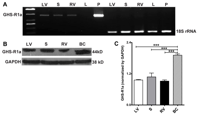
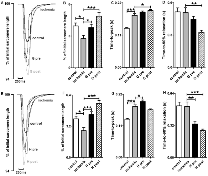
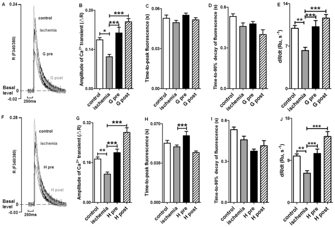
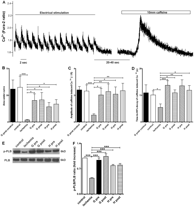
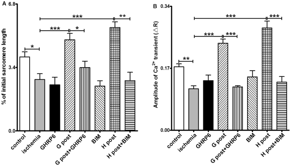
Methodology/principal findings: Isolated hearts from adult male mice underwent 20 min global ischemia and 30 min reperfusion using a Langendorff apparatus. Ghrelin (10 nM) or hexarelin (1 nM) was introduced into the perfusion system either 10 min before or after ischemia, termed pre- and post-treatments. In freshly isolated cardiomyocytes from these hearts, single cell shortening, intracellular calcium ([Ca(2+)](i)) transients and caffeine-releasable sarcoplasmic reticulum (SR) Ca(2+) were measured. In addition, RT-PCR and Western blots were used to examine the expression level of GHS receptor type 1a (GHS-R1a), and phosphorylated phospholamban (p-PLB), respectively. Ghrelin and hexarelin pre- or post-treatments prevented the significant reduction in the cell shortening, [Ca(2+)](i) transient amplitude and caffeine-releasable SR Ca(2+) content after I/R through recovery of p-PLB. GHS-R1a antagonists, [D-Lys3]-GHRP-6 (200 nM) and BIM28163 (100 nM), completely blocked the effects of GHS on both cell shortening and [Ca(2+)](i) transients.

Conclusion/significance: Through activation of GHS-R1a, ghrelin and hexarelin produced a positive inotropic effect on ischemic cardiomyocytes and protected them from I/R injury probably by protecting or recovering p-PLB (and therefore SR Ca(2+) content) to allow the maintenance or recovery of normal cardiac contractility. These observations provide supporting evidence for the potential therapeutic application of ghrelin and hexarelin in patients with cardiac I/R injury.


Conflict of interest statement

Competing Interests: The authors have declared that no competing interests exist.

Figures

Figure 1
Figure 1. mRNA (A) and protein (B and C) expression of GHS-R1a in mouse heart.
(A) Liver (L) and pituitary gland (P) were used as the negative and positive controls respectively. Mouse 18s rRNA was chosen as an internal control. (B) A breast cancer (BC) cell line was used as the positive control. (C) n  =  3 for Western blots, data are shown as means ± S.E.M. and analyzed by One-way ANOVA with Tukey’s post hoc test. ***P < 0.001 vs BC group. GHS-R1a mRNA and protein are expressed in the left ventricle (LV), septum (S) and right ventricle (RV) of the mouse heart.
Figure 2
Figure 2. Effects of GHS on the contractile properties of mouse cardiomyocytes exposed to ischemia/reperfusion.
(A) and (E) are representative superimposed traces of sarcomere shortening after ghrelin (G) or hexarelin (H) pre-treatment (pre) and post-treatment (post). Both hexarelin and ghrelin improved the reduction in sarcomere shortening (B and F), but not the prolonged time-to-peak shortening (C and G). The time-to-peak shortening was further prolonged in G post- and H pre-treatment groups. The time for relaxation (D and H) was shortened in G post-, H pre- and post-treatment groups. n  =  99, 84, 84, 95, 106 and 100 cells/3 mice in control, ischemia, G pre, G post, H pre and H post groups, respectively. Data are shown as means ± S.E.M. and analyzed by One-way ANOVA with Tukey’s post hoc test. *P < 0.05, ** P < 0.01, *** P < 0.001 vs ischemic group.
Figure 3
Figure 3. Effects of GHS on [Ca2+]i transients in cardiomyocytes exposed to ischemia?reperfusion.
(A) and (F) are representative superimposed traces of [Ca2+]i transient after ghrelin (G) or hexarelin (H) pre-treatment (pre) and post-treatments (post). R represents the emission fluorescence ratio of fura-2 from excitation at 340 and 380nm, and Ru represents the ratio unit. Both hexarelin and ghrelin improved the reduced amplitude of [Ca2+]i transients (B and G). The time-to-peak fluorescence was prolonged in ghrelin and hexarelin pre-treatment (C and H) groups, but the decreased rate of rise of [Ca2+]i transients after ischemia was reversed to control level by all H and G pre- and post-treatments (E and J). Ghrelin and hexarelin had no effect on the time-to-90% decay of [Ca2+]i transient (D and I). n  =  72, 80, 90, 103, 63 and 66 cells/3 mice in control, ischemia, G pre, G post, H pre and H post groups, respectively. Data are shown as means ± S.E.M. and analyzed by One-way ANOVA with Tukey’s post hoc test. *P < 0.05, **P < 0.01, *** P < 0.001 vs ischemic group.
Figure 4
Figure 4. Effects of GHS on sarcoplasmic reticulum (SR) Ca2+ content and phospho-phospholamban (p-PLB)/phospholamban (PLB) expression.
(A) Illustration of the SR Ca2+ content measurement protocol (data from cardiomyocytes exposed to ischemia/reperfusion). R represents the emission fluorescence ratio of fura-2 from excitation at 340 and 380nm. Cardiomyocytes were perfused with Tyrode solution containing 1.5 mM CaCl2 and paced at 0.5 Hz for at least 30 s. 10 mM caffeine was then added to induce SR Ca2+ release. SR Ca2+ content, as determined by both (B) Area under curve and (C) amplitude of the caffeine-induced Ca2+ release, significantly decreased after 20 min ischemia compared with the control group. Ghrelin (G) or hexarelin (H) pre-treatment (pre) and post-treatment (post) significantly increased the SR Ca2+ content after 20 min ischemia (B and C), but introduction of ghrelin into the perfusion system at 40 min and lasting for 10 min (G post control) had no effect on the SR Ca2+ content of the cells isolated from the normal perfused heart. (D) The time-to-90% decay of caffeine-induced increases in [Ca2+]i mainly reflect the Ca2+ clearance ability of the Na+/Ca2+ exchanger (NCX). n  =  18, 44, 44, 33, 38, 41 and 38 cells/3 mice in G post control, control, ischemia, G pre, G post, H pre and H post, respectively. (E) Representative western blots of the total phospholamban (PLB) and the phosphorylated PLB (p-PLB) in 6 groups and (F) the densitometric quantification of ratio of p-PLB/PLB (expressed as fold increase relative to control). n  =  5 mice in each group. Data are shown as means ± S.E.M. and analyzed by one-way ANOVA with Tukey’s post hoc test. *P < 0.05, **P < 0.01, *** P < 0.001 vs ischemic group.
Figure 5
Figure 5. Role of GHS-R1a in the cardiac effects of hexarelin (H) and ghrelin (G).
R represents the emission fluorescence ratio of fura-2 from excitation at 340 and 380nm. The GHS-R1a antagonists, [D-Lys3]-GHRP-6 (GHRP6, 200 nM) and BIM28163 (BIM,100 nM), completely blocked the effects of 1 nM hexarelin (H) and 10 nM ghrelin (G) post-treatment (post) on sarcomere shortening (A) and [Ca2+]i transients (B). These peptides alone did not produce any noticeable change in sarcomere shortening (A) and [Ca2+]i transients (B). For sarcomere shortening experiments, n  =  99, 84, 52, 95, 46,100, 50 and 61 in control, ischemic, GHRP6, G post, G post +GHRP6, BIM, H post and H post+BIM groups, respectively. For [Ca2+]i transient experiments, n  =  72, 80, 55, 103, 69, 60, 66 and 68 in control, ischemic, GHRP6, G post, G post +GHRP6, BIM, H post and H post+BIM groups, respectively. Data were analyzed by one-way ANOVA with Tukey's post hoc test, and expressed as means ± S.E.M. *P < 0.05, ** P < 0.01, *** P < 0.001.

Similar articles

Cited by

References

    1. Ostadal B, Kolář F. Cardiac ischemia: from injury to protection: Springer. 1999.
    1. Murphy E, Steenbergen C. Mechanisms underlying acute protection from cardiac ischemia-reperfusion injury. Physiol Rev. 2008;88:581–609. - PMC - PubMed
    1. Headrick JP, Peart J, Hack B, Flood A, Matherne GP. Functional properties and responses to ischaemia-reperfusion in Langendorff perfused mouse heart. Exp Physiol. 2001;86:703–716. - PubMed
    1. Furman E, Acad BA, Sonn J, Raul A, Kedem J. Effect of global vs regional ischaemia upon myocardial contractility and oxygen balance. Cardiovasc Res. 1985;19:606–612. - PubMed
    1. Chambers DJ, Fallouh HB. Cardioplegia and cardiac surgery: pharmacological arrest and cardioprotection during global ischemia and reperfusion. Pharmacol Ther. 2010;127:41–52. - PubMed

Publication types

MeSH terms