Spontaneous aortic rupture in a patient with neurofibromatosis type 1
- PMID: 22493769
- PMCID: PMC3319782
- DOI: 10.4174/jkss.2012.82.4.261
Spontaneous aortic rupture in a patient with neurofibromatosis type 1
Abstract
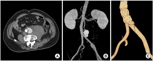
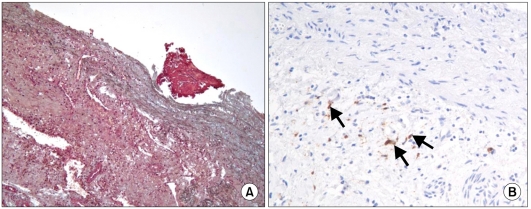
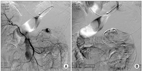
Neurofibromatosis type I (NF-1) is a rare autosomal dominant genetic disorder occurring in 1 in 3,000 individuals. Vasculopathy is a rarely reported finding in patients with NF-1. Here, we report a case of recurrent aortic pseudoaneurysm after endovascular aneurysm repair in a 49-year-old male patient with NF-1. On the sixth postoperative day following a successful open surgical repair of an aortic pseudoaneurysm, he developed hemoperitoneum due to a delayed rupture of the mesenteric artery branch. This was treated with endovascular coil embolization. We report the clinical features and histologic findings of this rare vascular disorder with a review of the relevant literature.
Keywords: Aortic aneurysm; Aortic rupture; Neurofibromatosis 1.
Conflict of interest statement
No potential conflict of interest relevant to this article was reported.
Figures
References
-
- Friedman JM, Arbiser J, Epstein JA, Gutmann DH, Huot SJ, Lin AE, et al. Cardiovascular disease in neurofibromatosis 1: report of the NF1 Cardiovascular Task Force. Genet Med. 2002;4:105–111. - PubMed
-
- National Institutes of Health Consensus Development Conference Statement: neurofibromatosis. Bethesda, Md., USA, July 13-15, 1987. Neurofibromatosis. 1988;1:172–178. - PubMed
-
- Shimizu T, Yamazaki Y, Tomoe H, Nishino S, Toma H, Shibata T, et al. Giant retroperitoneal hematoma in a patient with von Recklinghausen's disease. Nihon Hinyokika Gakkai Zasshi. 1998;89:846–849. - PubMed
-
- Chew DK, Muto PM, Gordon JK, Straceski AJ, Donaldson MC. Spontaneous aortic dissection and rupture in a patient with neurofibromatosis. J Vasc Surg. 2001;34:364–366. - PubMed
-
- Hines GL, Lefkowitz L, Mohtashemi M. Infrarenal aortic rupture secondary to neurofibromatosis. Ann Vasc Surg. 2002;16:784–786. - PubMed
Publication types
LinkOut - more resources
Full Text Sources
Research Materials
Miscellaneous