. 2012 Apr 11:12:20.
doi: 10.1186/1471-2377-12-20.

Dexmedetomidine is neuroprotective in an in vitro model for traumatic brain injury

Affiliations

Dexmedetomidine is neuroprotective in an in vitro model for traumatic brain injury

Marc Schoeler et al. BMC Neurol. .

Abstract

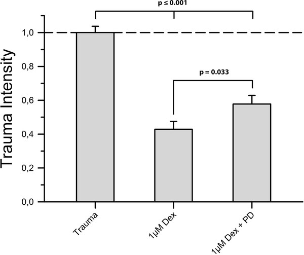
Background: The α2-adrenoreceptor agonist dexmedetomidine is known to provide neuroprotection under ischemic conditions. In this study we investigated whether dexmedetomidine has a protective effect in an in vitro model for traumatic brain injury.

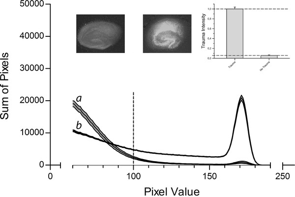
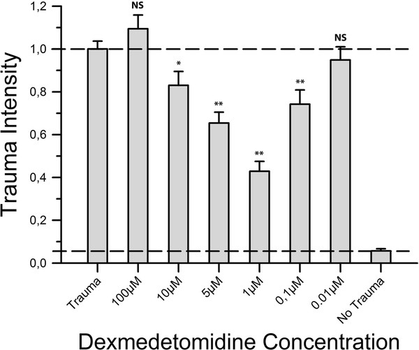
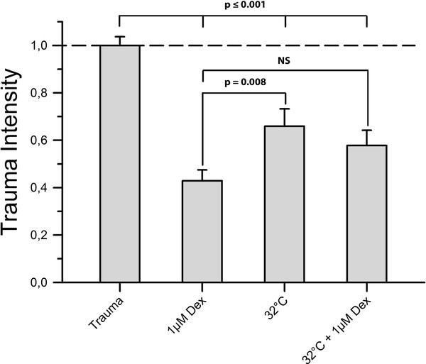
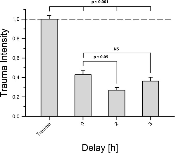
Methods: Organotypic hippocampal slice cultures were subjected to a focal mechanical trauma and then exposed to varying concentrations of dexmedetomidine. After 72 h cell injury was assessed using propidium iodide. In addition, the effects of delayed dexmedetomidine application, of hypothermia and canonical signalling pathway inhibitors were examined.

Results: Dexmedetomidine showed a protective effect on traumatically injured hippocampal cells with a maximum effect at a dosage of 1 μM. This effect was partially reversed by the simultaneous administration of the ERK inhibitor PD98059.

Conclusion: In this TBI model dexmedetomidine had a significant neuroprotective effect. Our results indicate that activation of ERK might be involved in mediating this effect.


Figures

Figure 1
Figure 1
Control Data. Slices were cultivated for 14 days before being subjected to TBI introduced by a stylus dropped onto the CA1 region. Slices were then incubated in an atmosphere of 95% air and 5% CO2. Negative control group slices were treated the same way, except for the trauma. After 72 h trauma intensity was assessed by fluorescence imaging and pixel based analysis. The fluorescence intensity corresponded to the trauma intensity of each pixel and was broken down to a value between 0 and 255. The histogram shows the distribution of pixels for the different fluorescence intensities for the negative control group (n = 35, labelled a) and the slices of the positive control group (n = 185, labelled b). The middle line represents the mean for each fluorescence value; the upper and lower lines represent the upper and lower boundaries of the SEM. The interrupted vertical line is the applied threshold at a grey scale value of 100. The sum of all pixels beyond this threshold was calculated for each group and served as a measure for trauma intensity. Above the graph, example images for the negative control group (left) and positive control group (right) are shown. The insert in the upper right corner represents the trauma levels of both control groups normalized to the positive control group.
Figure 2
Figure 2
Neuroprotective effect of dexmedetomidine. After trauma, slices were exposed to varying concentrations of dexmedetomidine. Following 72 h of incubation fluorescence images were taken and analysed. For each group an average of 52 slices with a minimum of 39 slices was used. Trauma intensities of the different groups are shown in relation to trauma intensity in the positive control group (n = 185). Trauma intensity was significantly lower in the groups 10 μM, 5 μM, 1 μM and 0.1 μM (*p ≤ 0.05, **p ≤ 0.001) when compared to the positive control group. In contrast, the groups 100 μM and 0.01 μM showed no statistical difference compared to the positive control group (p = 0.213 and p = 0.485, respectively).
Figure 3
Figure 3
Hypothermia. We subjected a group of slices (n = 49) to mild hypothermia of 32°C for 72 h after TBI to distinguish the observed dexmedetomidine protection from already determined neuroprotective treatment. Furthermore, we treated another group of slices (n = 68) simultaneously with both mild hypothermia and 1 μM dexmedetomidine to investigate a potential additive effect of both treatments. Figure 3 shows that treatment with mild hypothermia caused a significant reduction of trauma intensity when compared to the positive control group (p ≤ 0.001). However, this protective effect was significantly lower than protection provided by 1 μM dexmedetomidine (p = 0.008). Treatment with both hypothermia and 1 μM dexmedetomidine proved to be more effective in reducing trauma than hypothermia alone (p = 0.415) but showed no statistical difference compared to a treatment with 1 μM dexmedetomidine alone (p = 0.073).
Figure 4
Figure 4
Delayed application of dexmedetomidine. Here, we applied dexmedetomidine 2 and 3 h after TBI in a concentration of 1 μM. The two-hour delay group contained n = 24 slices and the three-hour delay group contained n = 31 slices. We found that both groups showed a level of trauma intensity significantly lower than the positive control group's (p ≤ 0.001). An application delayed by 2 h appeared to protect slices even better than immediate application of dexmedetomidine (p ≤ 0.05). After a delay of 3 h no statistical difference from an immediate application was observed (p = 0.335).
Figure 5
Figure 5
MEK-Inhibitor. To investigate whether the ERK1/2-pathway is involved in the mediation of dexmedetomidine's neuroprotective properties towards mechanically injured brain tissue we applied PD98059, inhibitor of ERK1/2's direct activator MEK1, in a 5 μM concentration along with 1 μM dexmedetomidine immediately after trauma. Here, n = 46 slices were subjected to the aforementioned conditions. As shown in Figure 5 trauma intensity was significantly higher in the inhibitor group than in the dexmedetomidine group (p = 0.033) but still appreciably lower than in the positive control group (p ≤ 0.001).

References

    1. Corrigan JD, Selassie AW, Orman JA. The epidemiology of traumatic brain injury. J Head Trauma Rehabil. 2010;25:72–80. doi: 10.1097/HTR.0b013e3181ccc8b4. - DOI - PubMed
    1. Selassie AW, Zaloshnja E, Langlois JA, Miller T, Jones P, Steiner C. Incidence of long-term disability following traumatic brain injury hospitalization, United States, 2003. J Head Trauma Rehabil. 2008;23:123–131. doi: 10.1097/01.HTR.0000314531.30401.39. - DOI - PubMed
    1. Thurman DJ, Alverson C, Dunn KA, Guerrero J, Sniezek JE. Traumatic brain injury in the United States: a public health perspective. J Head Trauma Rehabil. 1999;14:602–615. doi: 10.1097/00001199-199912000-00009. - DOI - PubMed
    1. Hoffman WE, Kochs E, Werner C, Thomas C, Albrecht RF. Dexmedetomidine improves neurologic outcome from incomplete ischemia in the rat. Reversal by the alpha 2-adrenergic antagonist atipamezole. Anesthesiology. 1991;75:328–332. doi: 10.1097/00000542-199108000-00022. - DOI - PubMed
    1. Dahmani S, Rouelle D, Gressens P, Mantz J. Characterization of the postconditioning effect of dexmedetomidine in mouse organotypic hippocampal slice cultures exposed to oxygen and glucose deprivation. Anesthesiology. 2010;112:373–383. doi: 10.1097/ALN.0b013e3181ca6982. - DOI - PubMed

MeSH terms

LinkOut - more resources