Review
. 2012 Jul-Aug;7(4):349-60.
doi: 10.1111/j.1747-0803.2012.00652.x. Epub 2012 Apr 12.

Finding the "PR-fect" solution: what is the best tool to measure fetal cardiac PR intervals for the detection and possible treatment of early conduction disease?

Affiliations
Review

Finding the "PR-fect" solution: what is the best tool to measure fetal cardiac PR intervals for the detection and possible treatment of early conduction disease?

Colin K L Phoon et al. Congenit Heart Dis. 2012 Jul-Aug.

Abstract

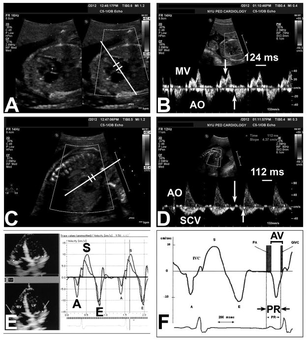
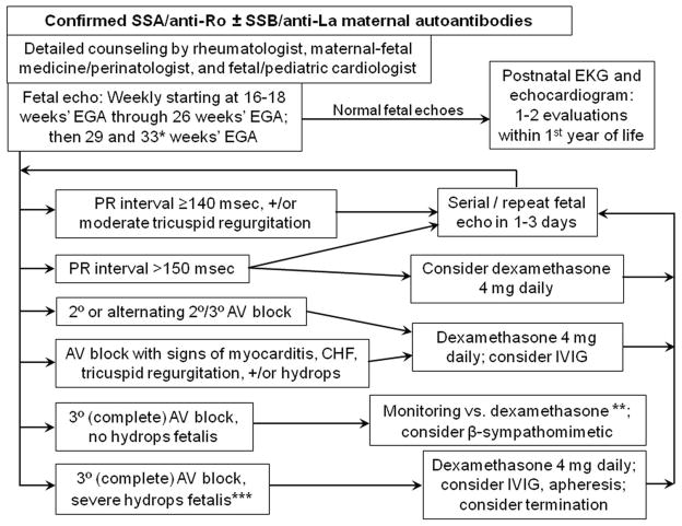
In the absence of structural heart disease, the great majority of cases with complete congenital heart block will be associated with the maternal autoantibodies directed to components of the SSA/Ro-SSB/La ribonucleoprotein complex. Usually presenting in fetal life before 26 weeks' gestation, once third-degree (complete) heart block develops, it is irreversible. Therefore, investigators over the past several years have attempted to predict which fetuses will be at risk for advanced conduction abnormalities by identifying a biomarker for less severe or incomplete disease, in this case, PR interval prolongation or first-degree atrioventricular block. In this state-of-the-art review, we critically analyze the various approaches to defining PR interval prolongation in the fetus, and then analyze several clinical trials that have attempted to address the question of whether complete heart block can be predicted and/or prevented. We find that, first and foremost, definitions of first-degree atrioventricular block vary but that the techniques themselves are all similarly valid and reliable. Nevertheless, the task of predicting those fetuses at risk, and who are therefore candidates for treatment, remains challenging. Of concern, despite anecdotal evidence, there is currently no conclusive proof that a prolonged PR interval predicts complete heart block.


Conflict of interest statement

CONFLICT OF INTEREST

Conflict of Interest: None declared.

Dr. Friedman is on the Speakers Bureau of and is a consultant for, MedImmune.

Figures

Figure 1
Figure 1. Ultrasound-based methods to measure mechanical surrogates of the electrocardiographic P-R interval in the fetus
A,B) Mitral-aorta pulsed-wave Doppler method; C,D) Superior caval vein-aorta pulsed-wave Doppler method; E,F) Fetal kinetocardiogram tissue Doppler method (with permission). A,B) The Mitral (MV)-aorta (AO) method relies on identification of the trans-mitral atrial inflow (A wave) and ejection out the left ventricular outflow tract. The Doppler pulsed-wave sample volume is placed near the mitral valve leaflet in the LV outflow tract (panel A), and measurement is made from the onset of the trans-mitral A wave to the onset of LV outflow tract ejection (panel B, arrows). In this case, the MV-AO P-R interval is 124 msec. C,D) The Superior caval vein (SCV)-aorta (AO) method interrogates both the caval and ascending aortic blood flow by pulsed-wave Doppler (panel C) and relies on the identification of onset of atrial “kick” reversal in the superior caval vein and aortic ejection (panel D, arrows). E,F [adapted from ref. , with permission]) The fetal kinetocardiogram relies on tissue Doppler imaging (panel E) and identification of atrial (late diastolic wall motion or A′) and ventricular motion (isovolumic ventricular contraction spike) to determine the “AV interval”, which mirrors the P-R interval (panel F).
Figure 2
Figure 2. Monitoring and management scheme for autoantibody-positive mothers
* = The original PRIDE protocol, to which we adhered in clinical practice until recently, called for biweekly monitoring from 26 through 34 weeks, in addition to the weekly scans from 16–26 weeks. Given the rarity of progression to atrioventricular block during this period, we now opt for more flexibility of monitoring during this time; as the diagram shows, we now have decreased the frequency of monitoring to twice between 26 and 34 weeks’ gestational age, once at about 29 weeks and once at about 33 weeks, if the fetus has been entirely well all along. Others have advocated close monitoring through approximately 35 weeks if there has been a previous child with CHB, which we agree is warranted. It should be noted that the definition of P-R interval prolongation will depend on the reference standards for the specific method used; values are shown for our laboratory using the Mitral-aorta Doppler method. ** = Third-degree (complete) atrioventricular block without fetal hydrops or other evidence of fetal compromise may be new-onset or may be established. For new-onset atrioventricular (AV) block (when sinus rhythm had been very recently observed), a trial of dexamethasone 4 mg daily is reasonable, in the slight chance that this may be reversed. β-sympathomimetic agents may also be considered in combination with dexamethasone, in efforts to increase the fetal heart rate. Otherwise, close (at least weekly) monitoring of fetal health, in close collaboration with maternal-fetal medicine/perinatology, is recommended. *** = In addition to fetal hydrops, evidence of myocarditis or cardiomyopathy, endocardial fibroelastosis, and/or significant tricuspid regurgitation, might also warrant aggressive therapy in an effort to prevent almost-certain fetal demise.

Similar articles

Cited by

References

    1. Buyon JP, Clancy RM, Friedman DM. Autoimmune associated congenital heart block: integration of clinical and research clues in the management of the maternal/foetal dyad at risk. J Intern Med. 2009;265:653–662. - PMC - PubMed
    1. Jaeggi ET, Silverman ED, Yoo S-J, Kingdom J. Is immune-mediated complete fetal atrioventricular block reversible by transplacental dexamethasone therapy? Ultrasound Obstet Gynecol. 2004;23:602–605. - PubMed
    1. Gordon PA. Congenital heart block: clinical features and therapeutic approaches. Lupus. 2007;16:642–646. - PubMed
    1. Bruer JMPJ, Kapusta L, Stoutenbeek P, Visser GHA, Van den Berg P, Meijboom EJ. Isolated congenital complete atrioventricular block diagnosed in utero: Natural history and outcome. J Mat Fetal Neonat Med. 2008;21:469–476. - PubMed
    1. Mevorach D, Elchalal U, Rein AJJT. Prevention of complete heart block in children of mothers with anti-SSA/Ro and anti-SSB/La autoantibodies: detection and treatment of first-degree atrioventricular block. Curr Opin Rheumatol. 2009;21:478–482. - PubMed

Publication types

Supplementary concepts