. 2012 Jul 18;18(1):825-33.
doi: 10.2119/molmed.2012.00005.

Extracellular histones inhibit efferocytosis

Affiliations

Extracellular histones inhibit efferocytosis

Arnaud Friggeri et al. Mol Med. .

Abstract

The uptake and clearance of apoptotic cells by macrophages and other phagocytic cells, a process called efferocytosis, is a major component in the resolution of inflammation. Increased concentrations of extracellular histones are found during acute inflammatory states and appear to contribute to organ system dysfunction and mortality. In these studies, we examined the potential role of histones in modulating efferocytosis. We found that phagocytosis of apoptotic neutrophils or thymocytes by macrophages was significantly diminished in the presence of histones H3 or H4, but not histone H1. Histone H3 demonstrated direct binding to macrophages, an effect that was diminished by preincubation of macrophages with the opsonins growth arrest-specific gene 6 (Gas6) and milk fat globule-epidermal growth factor (EGF) 8 (MFG-E8). Incubation of histone H3 with soluble α(v)β₅ integrin and Mer, but not with α(v)β₃, diminished its binding to macrophages. Phagocytosis of apoptotic cells by alveolar macrophages in vivo was diminished in the presence of histone H3. Incubation of histone H3 with activated protein C, a treatment that degrades histones, abrogated its inhibitory effects on efferocytosis under both in vitro and in vivo conditions. The present studies demonstrate that histones have inhibitory effects on efferocytosis, suggesting a new mechanism by which extracellular histones contribute to acute inflammatory processes and tissue injury.


Figures

Figure 1
Figure 1
Histones inhibit efferocytosis. Peritoneal macrophages were incubated with apoptotic neutrophils (A) or apoptotic thymocytes (B) in medium containing 10 μg/mL BSA (control) or histone H3 at increasing concentrations (1, 5 or 10 μg/mL) for 60 min. Efferocytosis assays were then performed as described in Materials and Methods. The percentage of macrophages that phagocytosed apoptotic cells for the control group is shown above the bar. Fold changes were calculated by dividing the percentage of macrophages that phagocytosed apoptotic cells for the experiment groups by that of the control groups. (C) Peritoneal macrophages were exposed to 10 μg/mL BSA (control), histone H3 (1, 5 or 10 μg/mL) or medium for 60 min and then levels of LDH (optical density [O.D.]) in the culture supernatants were determined. (D) Histones H3 and H4, but not histone H1, inhibit efferocytosis. Macrophages were incubated for 60 min with apoptotic thymocytes in media containing BSA (control) or histones H1, H3 or H4 (10 μg/mL), and then efferocytosis assays were performed. *p < 0.05, **p < 0.01, and ***p < 0.001 compared with the control group. The percentage of macrophages that phagocytosed apoptotic cells for the control group is shown above the bar. Fold changes were calculated by dividing the percentage of macrophages that phagocytosed apoptotic cells for the experiment groups by that of the control groups.
Figure 2
Figure 2
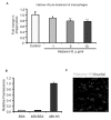
Histone H3 inhibits efferocytosis by binding to macrophages. (A) Macrophages were preincubated with BSA (10 μg/mL) or increasing doses of histone H3 (1, 5 and 10 μg/mL) for 1 h. The macrophages were then washed with fresh medium to remove unbound proteins, and apoptotic thymocytes were added for 60 min, after which efferocytosis assays were performed. (B) Macrophages were incubated with BSA (control), Chromeo 488–conjugated BSA (BSA-488) or Chromeo 488–conjugated histone H3 (H3-488) (5 μg/mL) for 1 h. The cells were then washed three times with PBS to remove unbound proteins. The quantities of proteins bound to macrophages were determined by a fluorescent plate reader. (C) Peritoneal macrophages were plated on coverslips and incubated with 5 μg/mL Chromeo 488–conjugated histone H3 (Histone H3-488) for 1 h. The macrophages were then washed three times with PBS, and confocal fluorescent microscopy analysis was performed to determine bound histone H3. DAPI was used to stain nuclei. **p < 0.01 compared with the control group.
Figure 3
Figure 3
Binding of histone H3 to macrophages is diminished by the opsonins Gas6 and MFG-E8. Macrophages were incubated with BSA (control), Gas6 or MFG-E8 (5 μg/mL) for 1 h and then washed three times, followed by incubation with Chromeo 488–conjugated histone H3 (H3-488) or Chromeo 488–conjugated BSA (BSA-488) (5 μg/mL) for 1 h. The cells were then washed again with PBS to remove unbound proteins. The quantities of proteins bound to macrophages were determined by a fluorescent plate reader. *p < 0.05 and ***p < 0.001 compared with the control group.
Figure 4
Figure 4
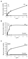
Histone H3 binds to the αvβ5 integrin and to the Mer receptor but not to the αvβ3 integrin. The 96-well plates were precoated with histone H3 or BSA (1 μg/mL) in PBS. The plates were then washed three times with PBS and blocked with PBS containing 1% BSA for 1 h. After removing the blocking solution, the plates were incubated with increasing doses (0, 0.1, 0.5, 1 and 5 μg/mL) of recombinant mouse Mer (A), αvβ5 (B) or αvβ3 (C) dissolved in PBS for 1 h, followed by washing with PBS containing 0.05% Tween 20. Protein bound to the wells was quantified by solid-phase ELISA as described in Materials and Methods. A450, absorbance at 450 nm.
Figure 5
Figure 5
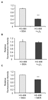
Binding of histone H3 to macrophages is diminished by the integrin αvβ5 and Mer, but not by the αvβ3 integrin. Chromeo 488–conjugated histone H3 (H3-488) (5 μg/mL) was incubated with BSA or Mer (A), integrin αvβ3 (B) or integrin αvβ5 (C) (5 μg/mL) for 30 min in RPMI-1640. The mixture of the proteins was then added to macrophages and incubated for 1 h, after which the cells were washed three times with PBS and the quantities of bound protein were determined by a fluorescent plate reader. ***p < 0.001 compared with the 488-H3 + BSA group.
Figure 6
Figure 6
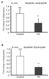
Histone H3 inhibits efferocytosis in vivo. Mice (three in each group) were exposed to the intratracheal administration of 10 × 106 apoptotic neutrophils (A) or apoptotic thymocytes (B) resuspended in 50 μL PBS containing 5 μg BSA (control) or histone H3, and bronchoalveolar lavages were collected 2 h later. Cytospin slides were prepared from the bronchoalveolar lavage fluid, and phagocytosis was determined by microscopy (A) or by flow cytometry (B). **p < 0.05 compared with the BSA group. The percentage of macrophages that phagocytosed apoptotic cells for the control group is shown above the bar.
Figure 7
Figure 7
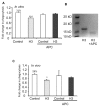
APC abrogates the inhibitory effects of histone H3 on efferocytosis in vitro and in vivo. (A) Macrophages were incubated for 1 h with apoptotic thymocytes in the presence of 5 μg/mL BSA (control), histone H3, BSA that was preincubated with 200 nmol/L APC or histone H3 that was preincubated with 200 nmol/L APC, after which efferocytosis assays were performed. (B) Histone H3 and histone H3 preincubated with 200 nmol/L APC were resolved by SDS-PAGE (sodium dodecyl sulfate–polyacrylamide gel electrophoresis). The gel was stained with Coomassie Blue R-250 to visualize histone H3 and cleaved histone H3. (C) Apoptotic thymocytes were resuspended in 50 μL PBS containing 5 μg BSA (control), histone H3, BSA that was preincubated with 200 nmol/L APC or histone H3 that was preincubated with 200 nmol/L APC and then administered intratracheally for in vivo efferocytosis assays. *p < 0.05 and ***p < 0.001 in comparison with the BSA group. The percentage of macrophages that phagocytosed apoptotic cells for the control group is shown above the bar.

Similar articles

Cited by

References

    1. Vandivier RW, Henson PM, Douglas IS. Burying the dead: the impact of failed apoptotic cell removal (efferocytosis) on chronic inflammatory lung disease. Chest. 2006;129:1673–82. - PubMed
    1. Blasi F, Sidenius N. Efferocytosis: another function of uPAR. Blood. 2009;114:752–3. - PubMed
    1. Armstrong A, Ravichandran KS. Phosphatidylserine receptors: what is the new RAGE? EMBO Rep. 2011;12:287–8. - PMC - PubMed
    1. Elliott MR, Ravichandran KS. Clearance of apoptotic cells: implications in health and disease. J Cell Biol. 2010;189:1059–70. - PMC - PubMed
    1. Ravichandran KS. Find-me and eat-me signals in apoptotic cell clearance: progress and conundrums. J Exp Med. 2010;207:1807–17. - PMC - PubMed

Publication types

MeSH terms

LinkOut - more resources