

Circulating IL-15 exists as heterodimeric complex with soluble IL-15Rα in human and mouse serum
- PMID: 22496150
- PMCID: PMC3390963
- DOI: 10.1182/blood-2011-10-384362
Circulating IL-15 exists as heterodimeric complex with soluble IL-15Rα in human and mouse serum
Abstract
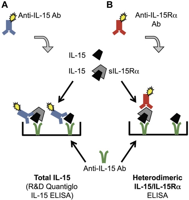
IL-15 is an important cytokine for the function of the immune system, but the form(s) of IL-15 produced in the human body are not fully characterized. Coexpression of the single-chain IL-15 and the IL-15 receptor alpha (IL-15Rα) in the same cell allows for efficient production, surface display, and eventual cleavage and secretion of the bioactive IL-15/IL-15Rα heterodimer in vivo, whereas the single-chain IL-15 is poorly secreted and unstable. This observation led to the hypothesis that IL-15 is produced and secreted only as a heterodimer with IL-15Rα. We purified human IL-15/IL-15Rα complexes from overproducing human cell lines and developed an ELISA specifically measuring the heterodimeric form of IL-15. Analysis of sera from melanoma patients after lymphodepletion revealed the presence of circulating IL-15/IL-15Rα complexes in amounts similar to the total IL-15 quantified by a commercial IL-15 ELISA that detects both the single-chain and the heterodimeric forms of the cytokine. Therefore, in lymphodepleted cancer patients, the serum IL-15 is exclusively present in its heterodimeric form. Analysis of the form of IL-15 present in either normal or lymphodepleted mice agrees with the human data. These results have important implications for development of assays and materials for clinical applications of IL-15.
Figures
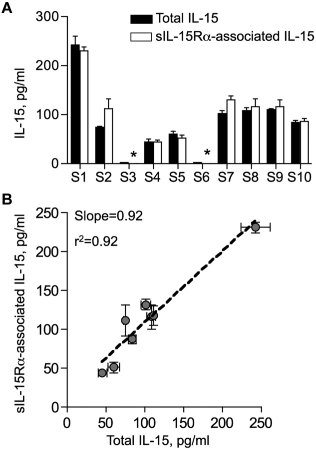
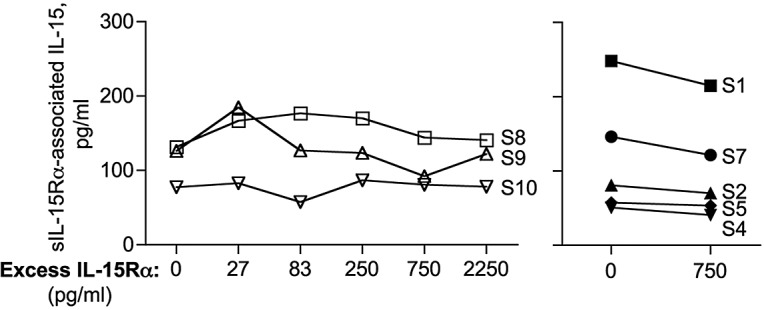
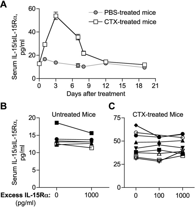
 ) and on CTX treatment (□). Mice were bled at different time points (days 1-19) after CTX or placebo administration. Circulating IL-15/IL-15Rα was assessed by ELISA (eBioscience) and reported as mean ± SEM of 6 to 33 mice per time point. (B-C) Serum samples from 6 untreated (B) and 9 CTX-treated (day 3 after treatment; C) mice were preincubated with exogenous recombinant mouse IL-15Rα-Fc (100-1000 pg/mL, as indicated) to allow the in vitro formation of IL-15/sIL-15Rα complexes with any free IL-15 potentially present in the sample. Serum samples were next evaluated for the amount of sIL-15Rα–associated IL-15 by ELISA.
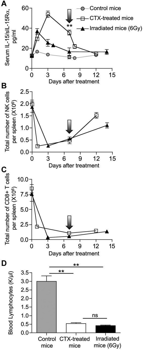 represents the average level of circulating mouse IL-15/IL-15Rα from 10 normal mice. **P = .007. (B-C) Mice were killed at the indicated time points after treatment, and spleens were collected for analysis. Total number of NK cells (B) and CD8+ T cells (C) per spleen over time was determined by flow cytometry. (D) Absolute count of lymphocytes in blood at day 7 after cytoreductive treatments. Three mice per group were analyzed. Hematologic profile on 10 untreated mice was also performed as control. **P < .01. ns indicates not significant.
Comment in
-
Soluble IL-15/IL-15Rα complexes in human serum.Blood. 2012 Jul 5;120(1):1-2. doi: 10.1182/blood-2012-04-425306. Blood. 2012. PMID: 22767572
References
-
- Grabstein KH, Eisenman J, Shanebeck K, et al. Cloning of a T cell growth factor that interacts with the beta chain of the interleukin-2 receptor. Science. 1994;264(5161):965–968. - PubMed
-
- Berard M, Brandt K, Bulfone-Paus S, Tough DF. IL-15 promotes the survival of naive and memory phenotype CD8+ T cells. J Immunol. 2003;170(10):5018–5026. - PubMed
-
- Ma A, Koka R, Burkett P. Diverse functions of IL-2, IL-15, and IL-7 in lymphoid homeostasis. Annu Rev Immunol. 2006;24:657–679. - PubMed
Publication types
MeSH terms
Substances
Grants and funding
LinkOut - more resources
Full Text Sources
Other Literature Sources
Molecular Biology Databases
