. 2012 Jul;60(7):550-9.
doi: 10.1369/0022155412441707. Epub 2012 Apr 11.

Sustained expression of osteopontin is closely associated with calcium deposits in the rat hippocampus after transient forebrain ischemia

Affiliations

Sustained expression of osteopontin is closely associated with calcium deposits in the rat hippocampus after transient forebrain ischemia

Jang-Mi Park et al. J Histochem Cytochem. 2012 Jul.

Abstract

The present study was designed to evaluate the extent and topography of osteopontin (OPN) protein expression in the rat hippocampus 4 to 12 weeks following transient forebrain ischemia, and to compare OPN expression patterns with those of calcium deposits and astroglial and microglial reactions. Two patterns of OPN staining were recognized by light microscopy: 1) a diffuse pattern of tiny granular deposits throughout the CA1 region at 4 weeks after ischemia and 2) non-diffuse ovoid to round deposits, which formed conglomerates in the CA1 pyramidal cell layer over the chronic interval of 8 to 12 weeks. Immunogold-silver electron microscopy and electron probe microanalysis demonstrated that OPN deposits were indeed diverse types of calcium deposits, which were clearly delineated by profuse silver grains indicative of OPN expression. Intracellular OPN deposits were frequently observed within reactive astrocytes and neurons 4 weeks after ischemia but rarely at later times. By contrast, extracellular OPN deposits progressively increased in size and appeared to be gradually phagocytized by microglia or brain macrophages and some astrocytes over 8 to 12 weeks. These data indicate an interaction between OPN and calcium in the hippocampus in the chronic period after ischemia, suggesting that OPN binding to calcium deposits may be involved in scavenging mechanisms.


Conflict of interest statement

Declaration of Conflicting Interests: The authors declared no potential conflicts of interest with respect to the research, authorship, and/or publication of this article.

Figures

Figure 1.
Figure 1.
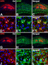
Spatiotemporal relationships of osteopontin (OPN) and alizarin red staining in the hippocampal CA1 region at 4 (A–C), 8 (D–F), and 12 weeks (G–L) after transient forebrain ischemia. (A–C) Tiny granular OPN deposits were diffusely distributed over the hippocampal CA1 region, which colocalized with amorphous to granule-like alizarin red Staining. pcl, the pyramidal cell layer; sr, stratum radiatum. (D−L) Over the chronic interval of 8 (1D) and 12 weeks (1G) after ischemia, non-diffuse, ovoid to round deposits of OPN immunostaining with variable size appeared to become enlarged and occupied the CA1 pyramidal cell layer, and their distribution closely matched that of alizarin red Staining. (J−L) Higher magnification views of the boxed areas in G to I, respectively. Note that the surfaces of OPN deposits were more intensely labeled than their cores. Cell nuclei appeared blue after DAPI staining. Scale bars A-I = 200µm; J-L = 50µm.
Figure 2.
Figure 2.
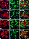
Relationship between osteopontin (OPN) protein expression and astroglial (A−F) and microglial (G−L) responses in the hippocampal CA1 region 4 weeks after transient forebrain ischemia. (A−F) Immunostaining for OPN and glial fibrillary acidic protein (GFAP) was observed throughout the CA1 hippocampus and revealed an overlapping regional distribution. (D−F) Higher magnification views of the CA1 region. Note that tiny OPN granules were observed within the cytoplasm of astrocytes with a hypertrophic appearance (asterisks in D−F). (G−L) Double labeling for OPN and Iba1 showed that some amoeboid brain macrophages contained dot-like labeled profiles. (J−L) Higher magnification views of the CA1 region. Note that tiny OPN granules (asterisks in J−L), which appeared to be localized in astrocytes, were not observed within brain macrophages. Cell nuclei appeared blue after DAPI staining. Scale bars A-C and G-I = 200µm; D-F and J-L = 20µm.
Figure 3.
Figure 3.
Relationship between osteopontin (OPN) protein and astroglial (A−C, G−I) and microglial (D−F, J−L) responses in the hippocampal CA1 region 8 (A−F) and 12 weeks (G−L) after transient forebrain ischemia. (A−C) Double labeling for OPN and glial fibrillary acidic protein (GFAP) showed that although some astrocytes showed tiny granular deposits of immunostaining (asterisks in A−C), most astrocytes were unlabeled or weakly labeled. Note that non-diffuse OPN-labeled deposits of variable sizes and shapes appeared to be located in the extracellular matrix. (D−F) Double labeling for OPN and Iba1 showed that brain macrophages were frequently located in close proximity to OPN deposits and sometimes contained these deposits within the cytoplasm (arrowheads in D−F). (G−I) Double labeling for OPN and GFAP showed that hypertrophic astroglial cytoplasm and processes were in close proximity to conglomerates of OPN-labeled deposits and sometimes encompassed them. Note that the cores of these deposits were devoid of specific labeling for OPN. (J−L) Double labeling for OPN and Iba1 showed that most clusters of OPN deposits were localized within cell bodies of brain macrophages. Cell nuclei appeared blue after DAPI staining. Scale bars A-L = 20µm.
Figure 4.
Figure 4.
Ultrastructural localization of osteopontin (OPN) by the immunogold-silver method in the CA1 region at 4 weeks following ischemia. (A, B) Silver grains indicative of OPN accumulated along the surface of electron-dense calcified deposits, which were irregular in shape and size and were located extracellularly surrounded by a large amount of cellular debris. Note that these deposits showed increased electron density at the periphery and sometimes contained degenerated mitochondria (arrowheads in B). (C, D) OPN-labeled deposits were observed within the soma (arrowheads in C) and dendrites (arrowheads in D) of degenerating neurons containing several lipid inclusions (arrows in C, D). nu, nucleus. (Inset in D) Higher magnification view of the boxed area in D. Note that silver grains were evenly localized within the intracellular deposit. (E, F) Some astrocytes contained lipid inclusions (arrow in E) and multiple OPN-labeled deposits, some of which were closely associated with glial filaments (f in F). (Inset in E) Higher magnification view of the boxed area in E. Note that silver grains were rather diffusely localized in the intracellular deposit. Scale bars A, C-E = 4µm; B and F = 1µm; D and E Insets = 0.5µm.
Figure 5.
Figure 5.
Ultrastructural localization of osteopontin (OPN) by the immunogold-silver method in the CA1 region at 8 weeks following ischemia. (A, B) Silver grains indicative of OPN accumulated along the surface of electron-dense calcified deposits, which were located intracellularly in dead neurons with few recognizable organelles. Note that these calcium deposits appeared to be fused to each other and much more electron dense than those at 4 weeks following ischemia. (Insets in A and B) Higher magnification views of the boxed areas in A and B, respectively. (C) The aggregated, extracellular calcium deposits (inset shows lower magnification) were delineated by profuse accumulations of silver grains. (D) A brain macrophage containing multiple calcium deposits with conspicuous enrichments of silver grains. Note that the silver grains also accumulated along the surface of aggregated extracellular calcium deposits (E in D). nu, nucleus. (E) A brain macrophage containing large clusters of calcium deposits with profuse silver grains. (F) A brain macrophage containing labeled deposits showed prominent features of phagocytic activity, such as lipophagosomes and lipid droplets (arrows in F). (Inset in F) Higher magnification view of the boxed area in F. Note silver grains in the engulfed deposits (arrowheads). Scale bars A, B, and C inset = 4µm; D and F = 2µm; B, C, E, and A and F Insets = 1µm.
Figure 6.
Figure 6.
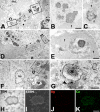
Ultrastructural localization of osteopontin (OPN) in association with reactive astrocytes (A−G) and elemental maps of OPN-labeled deposit (H−K) in the CA1 region at 8 weeks following ischemia. (A, D) Astrocytes showing extensive hypertrophy were frequently observed in close proximity to OPN-labeled calcium deposits. (B, C, E) Higher magnification views of the boxed areas in A and D, respectively. Note that astroglial processes were closely associated with OPN-labeled deposits (B) or completely encompassed them (C, E). Also note dense bundles of parallel-running glial filaments in astrocytes (f in B, C, E). nu, nucleus. (F, G) A reactive astrocyte containing an OPN-labeled deposit (asterisk in G) with a phagolysosome (arrowhead in G), providing evidence of phagocytosis by an astrocyte. A phagocytic brain macrophage (M) is observed in the vicinity. (G) Higher magnification view of the boxed area in F. f denotes bundles of glial filaments in astrocytic cytoplasm. (H−K) Transmission electron microscopy (TEM) and scanning TEM (STEM) images and corresponding compositional images of an identical ultrathin section. Electron probe microanalysis indicates the presence and the spatial distribution of silver (Ag, red) and calcium (Ca, green) within the labeled profile. Note that the prominent signals for calcium were observed within the labeled profile and that silver signals corresponded with silver grains for OPN. Scale bars A, D, F = 4µm; B, C, and G = 1µm; E, H-K = 0.5µm.

References

    1. Agnew WF, Yuen TG, Bullara LA, Jacques D, Pudenz RH. 1979. Intracellular calcium deposition in brain following electrical stimulation. Neurol Res. 1:187–202 - PubMed
    1. al-Ali SY, al-Hussain SM. 1996. An ultrastructural study of the phagocytic activity of astrocytes in adult rat brain. J Anat. 188:257–262 - PMC - PubMed
    1. Bendel O, Bueters T, von Euler M, Ove Ogren S, Sandin J, von Euler G. 2005. Reappearance of hippocampal CA1 neurons after ischemia is associated with recovery of learning and memory. J Cereb Blood Flow Metab. 25:1586–1595 - PubMed
    1. Bonnekoh P, Kuroiwa T, Kloiber O, Hossmann K. 1992. Time profile of calcium accumulation in hippocampus, striatum and frontoparietal cortex after transient forebrain ischemia in the gerbil. Acta Neuropathol. 84:400–406 - PubMed
    1. Boskey AL, Maresca M, Ullrich W, Doty SB, Butler WT, Prince CW. 1993. Osteopontin-hydroxyapatite interactions in vitro: inhibition of hydroxyapatite formation and growth in a gelatin-gel. Bone Miner. 22:147–159 - PubMed

Publication types