. 2012 Jun;86(12):6815-24.
doi: 10.1128/JVI.00015-12. Epub 2012 Apr 11.

Genetic analysis of cytomegalovirus in malignant gliomas

Affiliations

Genetic analysis of cytomegalovirus in malignant gliomas

Bornali Bhattacharjee et al. J Virol. 2012 Jun.

Abstract

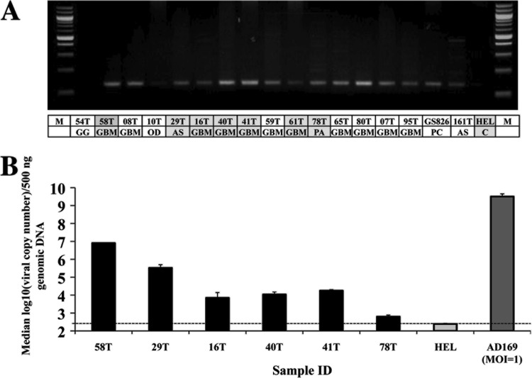
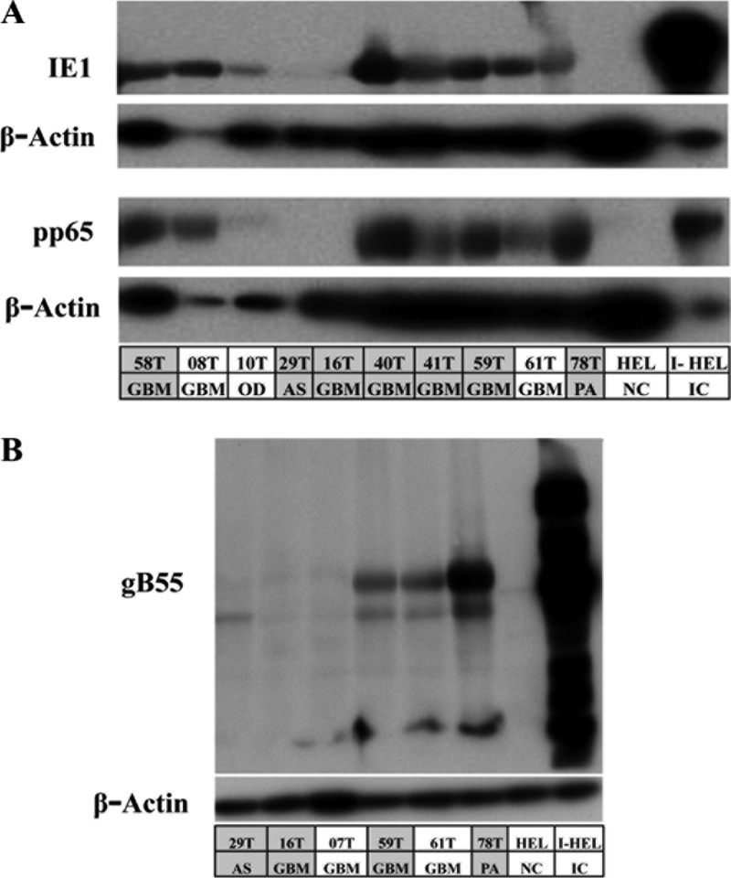
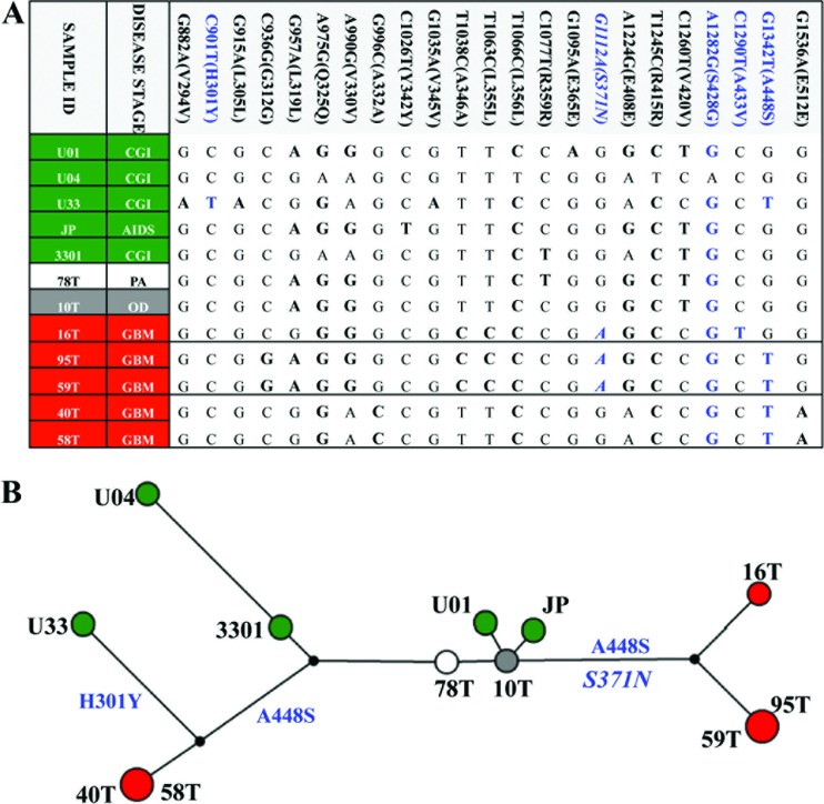
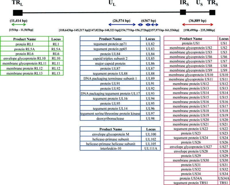
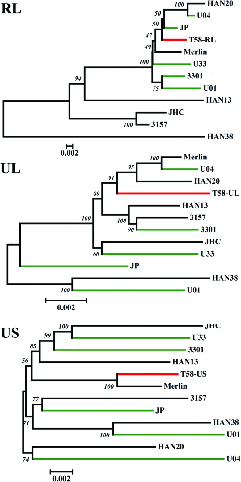
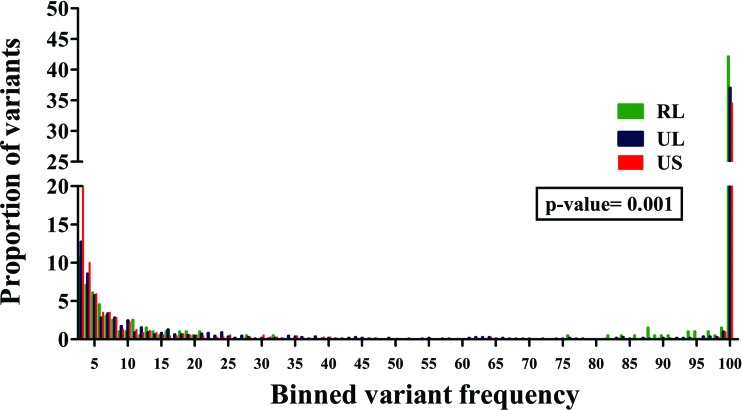
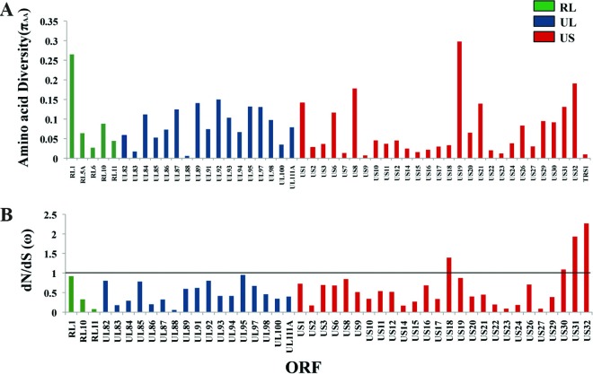
Human cytomegalovirus (HCMV) has been found in malignant gliomas at variable frequencies with efforts to date focused on characterizing the role(s) of single gene products in disease. Here, we reexamined the HCMV prevalence in malignant gliomas using different methods and began to dissect the genetics of HCMV in tumors. HCMV DNA was found in 16/17 (94%) tumor specimens. Viral DNA copy numbers were found to be low and variable, ranging from 10(2) to 10(6) copies/500 ng of total DNA. The tumor tissues had incongruences between viral DNA copy numbers and protein levels. However, nonlatent protein expression was detected in many tumors. The viral UL83 gene, encoding pp65, was found to segregate into five cancer-associated genotypes with a bias for amino acid changes in glioblastoma multiforme (GBM) in comparison to the low-grade tumors. Deep sequencing of a GBM-associated viral population resulted in 81,224 bp of genome coverage. Sequence analysis revealed the presence of intact open reading frames and higher numbers of high-frequency variations within the repeat long region compared to the unique long region, which harbors many core genes, and the unique short region (P = 0.001). This observation was in congruence with phylogenetic analyses across replication-competent viral strains in databases. The tumor-associated viral population was less variable (π = 0.1% and π(AA) = 0.08%) than that observed in other clinical infections. Moreover, 42/46 (91.3%) viral genes analyzed had dN/dS scores of <1, which is indicative of high amino acid sequence conservation. Taken together, these findings raise the possibility that replication-competent HCMV may exist in malignant gliomas.


Figures

Fig 1
Fig 1
Detection of HCMV DNA in tumor specimens. (A) Nested PCR products run on a 2% agarose gel. The amplicon is 144 bp in size. The first row in the table lists the sample identification, and the second row indicates the disease stage. M, 100-bp marker; GG, ganglioglioma; GBM, glioblastoma multiforme; OD, oligodendroma; AS, astrocytoma; PA, pilocytic astrocytoma; PC, primary culture of GBM tissue; C, uninfected HELs. The entries shaded in gray indicate patient specimens that were tested for viral copy number by subsequent real-time PCR. (B) Plot of median log10 (viral copy number) values from patient specimens. The dotted line denotes the lower limit of viral DNA detection.
Fig 2
Fig 2
Detection of IE, E, and L viral protein markers in gliomas. Immunoblots show the presence of viral proteins in patient specimens. The first row in the table lists the sample identification, and the second row indicates the disease stage. GBM, glioblastoma multiforme; OD, oligodendroma; AS, astrocytoma; PA, pilocytic astrocytoma. HEL, human embryonic lung fibroblasts; I-HEL, infected HEL cells (IC, infected cells). The entries shaded in gray are patient specimens that were tested for viral copy numbers in Fig. 1B. (A) Nine of twelve (75%) patient specimens were found to exhibit IE (IE1) and E (pp65) protein expression. (B) E/L (gB55) protein expression was found in the subset of tumors expressing IE and E proteins.
Fig 3
Fig 3
Viral pp65 genotype network of viral genotypes found in cancer specimens. (A) Genotypes were constructed from 23 variations. Nucleotides in boldface are variants relative to the Merlin reference genome, with nonsynonymous variants indicated in blue. Five genotypes were identified, and the sequence of each is shown in the table. An HLA-restricted polymorphism (G1112A) is indicated in italics. CGI, congenitical CMV infection. (B) A genotype network was inferred by the median-joining method with seven cancer-associated sequences using Merlin as the reference sequence. The size of each circle represents the frequencies of each genotype, with each color showing the disease stage of the patient as noted in panel A.
Fig 4
Fig 4
Schematic overview of the HCMV genomic regions included for deep sequencing. The black line is a schematic of the HCMV genome, which is organized into two unique regions—UL (unique long) and US (unique short)—flanked by three repeat regions: TRL, terminal repeat long; IRS, internal repeat short; and TRS, terminal repeat short. The arrows below the line indicate the regions amplified using overlapping primers. All of the nucleotide position numbers are given in reference to the Merlin genome. The numbers above the arrows are the total numbers of base pairs amplified. The numbers below the arrows indicate the nucleotide positions covered. The tables list the ORFs sequenced within each region. The RL region is colored green, the UL region is colored blue, and the US region is colored red.
Fig 5
Fig 5
The evolutionary relationship of the cancer-associated genome was inferred using the neighbor-joining method. Optimal unrooted trees with the sum of branch lengths for RL = 0.24579274, UL = 0.05801844, and US = 0.14616272 are shown. The percentages of replicate trees in which the associated strains clustered together in the bootstrap test (1,000 replicates) are shown next to the branches. The trees are drawn to scale, with branch lengths in the same units as those of the evolutionary distances used to infer the phylogenetic tree. The evolutionary distances are in the units of the number of base substitutions per site. The rate variation among sites was modeled with a gamma distribution (shape parameter = 1). The analyses involved 12 HCMV genomic sequences. The viral strains with green branches are from congenital infections sequenced previously in the laboratory (44). The 58T viral strain with red branches is the GBM-associated virus.
Fig 6
Fig 6
A significant difference was found in the frequency distribution of variants across the different regions of viral genomic sequences within a GBM specimen. A frequency distribution of all of the variants found within the 58T GBM specimen across the RL (green), UL (blue), and US (red) regions. Kruskal-Wallis testing revealed a significant difference among the distributions, with a P value of 0.001.
Fig 7
Fig 7
Deep sequencing revealed amino acid variability across the viral ORFs within the 58T GBM specimen. (A) Plot showing amino acid diversity (πAA) across different ORFs. (B) Plot of variable dN/dS (ω) values across different ORFs. The RL region is indicated in green, the UL region is indicated in blue, and the US region is indicated in red. The straight line denotes a cutoff of ω = 1 under an assumption of neutrality. ORFs containing only nonsynonymous changes were not included in the dN/dS analysis.

References

    1. Arnoult D, et al. 2004. Cytomegalovirus cell death suppressor vMIA blocks Bax- but not Bak-mediated apoptosis by binding and sequestering Bax at mitochondria. Proc. Natl. Acad. Sci. U. S. A. 101:7988–7993 - PMC - PubMed
    1. Asito AS, et al. 2010. Elevated anti-Zta IgG levels and EBV viral load are associated with site of tumor presentation in endemic Burkitt's lymphoma patients: a case control study. Infect. Agents Cancer 5:13. - PMC - PubMed
    1. Bandelt H-J, Forster P, Röhl A. 1999. Median-joining networks for inferring intraspecific phylogenies. Mol. Biol. Evol. 16:37–48 - PubMed
    1. Boccardo E, Villa LL. 2007. Viral origins of human cancer. Curr. Med. Chem. 14:2526–2539 - PubMed
    1. Cinatl J, Vogel JU, Kotchetkov R, Doerr WH. 2004. Oncomodulatory signals by regulatory proteins encoded by human cytomegalovirus: a novel role for viral infection in tumor progression. FEMS Microbiol. Rev. 28:59–77 - PubMed

Publication types